. 2006 Dec;188(24):8627-37.
doi: 10.1128/JB.00978-06. Epub 2006 Oct 6.

Mycoplasma genitalium P140 and P110 cytadhesins are reciprocally stabilized and required for cell adhesion and terminal-organelle development

Affiliations

Mycoplasma genitalium P140 and P110 cytadhesins are reciprocally stabilized and required for cell adhesion and terminal-organelle development

Raul Burgos et al. J Bacteriol. 2006 Dec.

Abstract

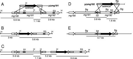
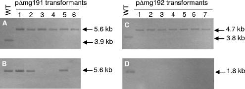
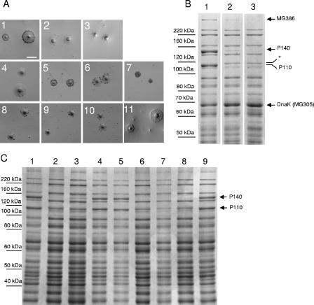
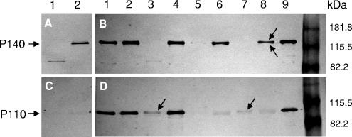
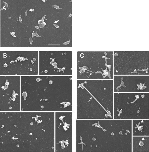
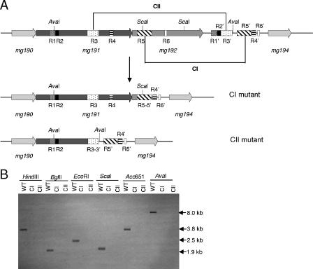
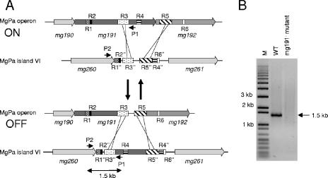
Mycoplasma genitalium is a human pathogen that mediates cell adhesion by a complex structure known as the attachment organelle. This structure is composed of cytadhesins and cytadherence-associated proteins, but few data are available about the specific role of these proteins in M. genitalium cytadherence. We have deleted by homologous recombination the mg191 and mg192 genes from the MgPa operon encoding the P140 and P110 cytadhesins. Molecular characterization of these mutants has revealed a reciprocal posttranslational stabilization between the two proteins. Loss of either P140 or P110 yields a hemadsorption-negative phenotype and correlates with decreased or increased levels of cytoskeleton-related proteins MG386 and DnaK, respectively. Scanning electron microscopy analysis reveals the absolute requirement of P140 and P110 for the proper development of the attachment organelle. The phenotype described for these mutants resembles that of the spontaneous class I and class II cytadherence-negative mutants [G. R. Mernaugh, S. F. Dallo, S. C. Holt, and J. B. Baseman, Clin. Infect. Dis. 17(Suppl. 1):S69-S78, 1993], whose genetic basis remained undetermined until now. Complementation assays and sequencing analysis demonstrate that class I and class II mutants are the consequence of large deletions affecting the mg192 and mg191-mg192 genes, respectively. These deletions originated from single-recombination events involving sequences of the MgPa operon and the MgPa island located immediately downstream. We also demonstrate the translocation of MgPa sequences to a particular MgPa island by double-crossover events. Based on these observations, we propose that in addition to being a source of antigenic variation, MgPa islands could be also involved in a general phase variation mechanism switching on and off, in a reversible or irreversible way, the adhesion properties of M. genitalium.


Figures

FIG. 1.
FIG. 1.
Homologous recombination at the MgPa operon in M. genitalium. (A and D) Schematic representation of the two possible crossover events of the pΔmg191 and pΔmg192 deletion constructs with the MgPa operon. (B and E) mg191 and mg192 genes, respectively, disrupted by double-crossover recombination. (C) mg191 gene disrupted by single-crossover recombination through the downstream flanking region. Hatched boxes represent flanking regions of the mg191 (lines from right to left) and mg192 (lines from left to right) genes in suicide plasmids pΔmg191 and pΔmg192, respectively. Black, tetM438 selectable marker; white, mg191 and mg192 genes; lines inside genes, probes S1, S2, and S3. The sizes of fragments resulting from HindIII (H) and BglII (Bg) digestions of the WT and each recombinant mutant DNA are also indicated.
FIG. 2.
FIG. 2.
Southern hybridization profiles of genomic DNAs from the M. genitalium WT strain and pΔmg191 and pΔmg192 transformants. A and B) HindIII digestions of genomic DNAs from the WT strain and pΔmg191 transformants hybridized with probes S3 and S1, respectively. (C and D) BglII digestions of DNAs from the WT strain and pΔmg192 transformants hybridized with probes S2 and S3, respectively. Probes S1, S2, and S3 are described in Fig. 1.
FIG. 3.
FIG. 3.
(A) Qualitative HA assessment of the different mutants and the WT strain. Panels 1 to 3, WT strain, mg192 mutant (clone 1), and mg191 mutant (clone 3), respectively. Panels 4 to 7, class I mutant, class I mutant plus pTnTetMG192, class I mutant plus pTnTetMG191, and class I mutant plus pTnTetMG191-192, respectively. Panels 8 to 11, class II mutant, class II mutant plus pTnTetMG192, class II mutant plus pTnTetMG191, and class II mutant plus pTnTetMG191-192, respectively. Bar, 100 μm. (B) SDS-8% PAGE protein profiles of the WT strain (lane 1) and the mg192 (clone 1) and mg191 (clone 3) mutants (lanes 2 and 3, respectively). Arrows show the differences observed between WT and mutant strains. *, new 115-kDa band. (C) SDS-8% PAGE protein profiles of the WT strain (lane 1), class I mutant (lane 2), class I mutant plus pTnTetMG191 (lane 3), class I mutant plus pTnTetMG192 (lane 4), class I mutant plus pTnTetMG191-192 (lane 5), class II mutant (lane 6), class II mutant plus pTnTetMG191 (lane 7), class II mutant plus pTnTetMG192 (lane 8), and class II mutant plus pTnTetMG191-192 (lane 9).
FIG. 4.
FIG. 4.
Western blot analyses of the different mutants and the WT strain. Blots were probed with an anti-P140 monoclonal antibody (A and B) or anti-P110 polyclonal antibody (C and D). In panels A and C, lanes 1 and 2 correspond to class II and class I mutants, respectively. In panels B and D, lanes 1 and 2 correspond to class II and class I mutants transformed with pTnTetMG191-192, respectively. Lanes 3 and 4, class II and class I mutants transformed with pTnTetMG192, respectively; lanes 5 and 6, are class II and class I mutants transformed with pTnTetMG191, respectively; lanes 7 and 8, mg191 mutant clone 4 and mg192 mutant clone 5, respectively; lane 9, WT strain. The arrows in panel B indicate the P140 doublet, while those in panel D indicate the presence of low quantities of P110.
FIG. 5.
FIG. 5.
Scanning electron microscopy analyses of the WT strain (A) and mg191 (B) and mg192 (C) mutants. Thick arrows indicate the terminal organelle, thin arrows show long filaments, and arrowheads show buds. All pictures are shown at the same magnification. Bar, 1 μm.
FIG. 6.
FIG. 6.
(A) Schematic representation of the DNA repetitive elements of the MgPa operon and MgPa island V located immediately downstream (bases 220000 to 234000 of the M. genitalium genome). A single-recombination event between the R5 and R5′ boxes and the R3 and R3′ boxes may generate class I and class II mutants, respectively. Base coordinates refer to accession number NC_000908 from the NCBI database. Boxes R1 to R6 refer to the DNA repetitive sequences of the MgPa operon, which are also found in the MgPa island (R1′ to R6′) located immediately downstream. CI, class I mutant; CII, class II mutant. (B) Southern hybridization profiles using probe S3 and genomic DNAs from the WT strain, class I mutant, and class II mutant.
FIG. 7.
FIG. 7.
Translocation of sequences from the MgPa operon to MgPa islands in the M. genitalium genome. (A) Schematic representation exemplifying how a double-recombination event between the R3-R3′ and R5-R5′ boxes translocates, in a reversible way, sequences from the MgPa operon (bases 220000 to 230000) to MgPa island VI (bases 310000 to 319000). Boxes R1 to R6 refer to the DNA repetitive sequences of the MgPa operon, which are also found in the MgPa island VI (R1′ to R6′) located 90 kb downstream. P1 and P2 indicate the position of the 3′P140XbaI and 5′mg260C-t primers, respectively. Base coordinates refer to accession number NC_000908 from the NCBI database. A number of additional double-recombination events are also possible but are not included in the drawing for clarity. (B) PCR amplification from M. genitalium WT and mg191 mutant DNAs, using primers P1 and P2. The 1.5-kb product results from a double-recombination event between R3 and downstream box R4, R5, or R6 of the MgPa operon and the corresponding boxes in the MgPa island VI. M, 1-kb plus DNA ladder marker.

References

    1. Baseman, J. B. 1993. The cytadhesins of Mycoplasma pneumoniae and M. genitalium. Subcell. Biochem. 20:243-259. - PubMed
    1. Baseman, J. B., M. Cagle, J. E. Korte, C. Herrera, W. G. Rasmussen, J. G. Baseman, R. Shain, and J. M. Piper. 2004. Diagnostic assessment of Mycoplasma genitalium in culture-positive women. J. Clin. Microbiol. 42:203-211. - PMC - PubMed
    1. Baseman, J. B., R. M. Cole, D. C. Krause, and D. K. Leith. 1982. Molecular basis for cytadsorption of Mycoplasma pneumoniae. J. Bacteriol. 151:1514-1522. - PMC - PubMed
    1. Baseman, J. B., S. F. Dallo, J. G. Tully, and D. L. Rose. 1988. Isolation and characterization of Mycoplasma genitalium strains from the human respiratory tract. J. Clin. Microbiol. 26:2266-2269. - PMC - PubMed
    1. Baseman, J. B., and J. G. Tully. 1997. Mycoplasmas: sophisticated, reemerging, and burdened by their notoriety. Emerg. Infect. Dis. 3:21-32. - PMC - PubMed

Publication types

MeSH terms

Substances

LinkOut - more resources