. 2006 Nov;174(3):1481-92.
doi: 10.1534/genetics.106.062794. Epub 2006 Oct 8.

Phylogenetic analysis of fungal centromere H3 proteins

Affiliations

Phylogenetic analysis of fungal centromere H3 proteins

Richard E Baker et al. Genetics. 2006 Nov.

Abstract

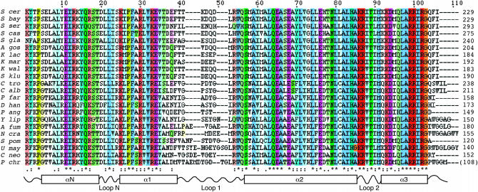
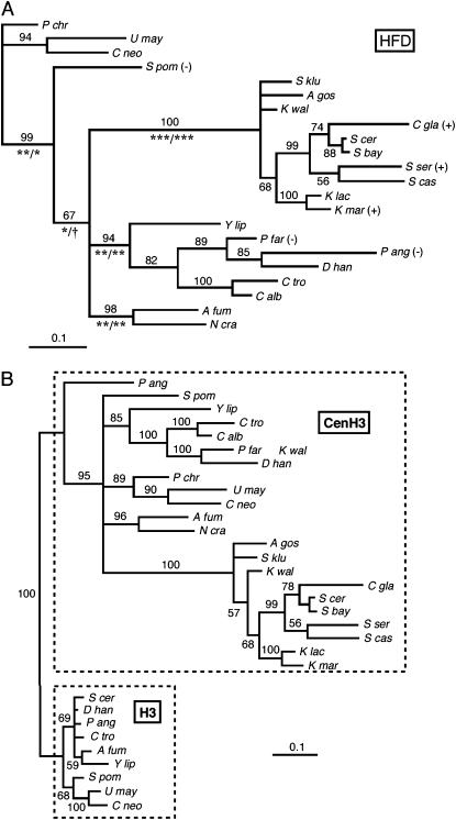
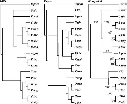
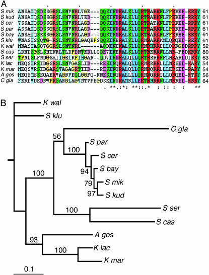
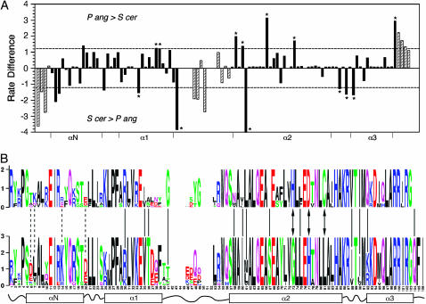
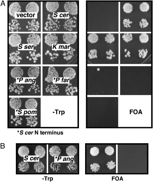
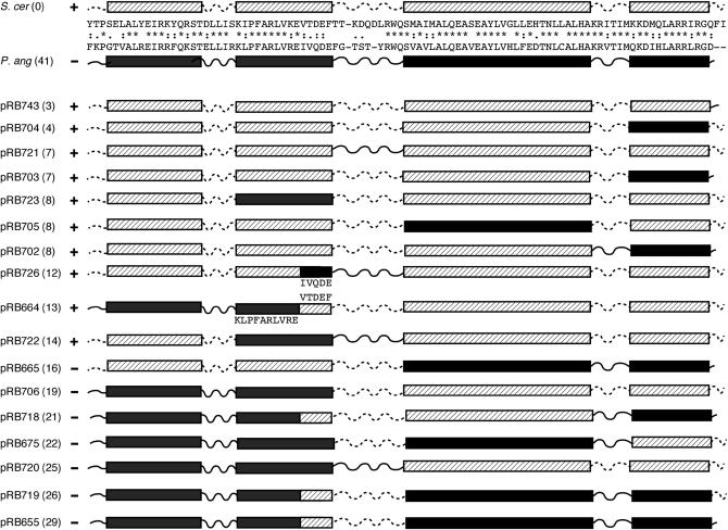
Centromere H3 proteins (CenH3's) are variants of histone H3 specialized for packaging centromere DNA. Unlike canonical H3, which is among the most conserved of eukaryotic proteins, CenH3's are rapidly evolving, raising questions about orthology and conservation of function across species. To gain insight on CenH3 evolution and function, a phylogenetic analysis was undertaken on CenH3 proteins drawn from a single, ancient lineage, the Fungi. Using maximum-likelihood methods, a credible phylogeny was derived for the conserved histone fold domain (HFD) of 25 fungal CenH3's. The collection consisted mostly of hemiascomycetous yeasts, but also included basidiomycetes, euascomycetes, and an archaeascomycete. The HFD phylogeny closely recapitulated known evolutionary relationships between the species, supporting CenH3 orthology. The fungal CenH3's lacked significant homology in their N termini except for those of the Saccharomyces/Kluyveromyces clade that all contained a region homologous to the essential N-terminal domain found in Saccharomyces cerevisiae Cse4. The ability of several heterologous CenH3's to function in S. cerevisiae was tested and found to correlate with evolutionary distance. Domain swapping between S. cerevisiae Cse4 and the noncomplementing Pichia angusta ortholog showed that species specificity could not be explained by the presence or absence of any recognized secondary structural element of the HFD.


Figures

F<sc>igure</sc> 1.—
Figure 1.—
HFD alignment. The alignment of 22 fungal CenH3 HFDs is shown in ClustalX format and coloring, with color intensity proportional to the conservation index. Secondary structural elements inferred from the three-dimensional structure of histone H3 are diagrammed schematically below the alignment. Four conserved positions N-terminal to the N-helix are included in the alignment. (The P. chrysosporium sequence is incomplete upstream of the region encoding the HFD.)
F<sc>igure</sc> 2.—
Figure 2.—
Fungal CenH3 and H3 phylogeny. (A) CenH3 HFD phylogeny. The phylogram shows the majority rule consensus tree produced by MrBayes, running a fixed-rate model with WAG rates (see materials and methods). Numbers above the branches are the percentage of times that branch was present in the posterior distribution of trees. Confidence intervals for the lengths of major branches were estimated under both fixed-rate and gamma substitution models. Symbols below the branches indicate, for each model, respectively, the level of significance at which the confidence interval excludes zero branch length (***, P = 0.001; **, P = 0.01; *, P = 0.05; †, P = 0.10). Plus (+) and minus (−) signs indicate whether or not the Cse4 ortholog complements Cse4 function in S. cerevisiae. (B) Nine fungal H3 sequences were added to the CenH3 HFD alignment, and a phylogeny was determined exactly as in A.
F<sc>igure</sc> 3.—
Figure 3.—
Tree topology tests. The left-hand cladogram shows the topology of the HFD tree with A. gossypii placed on the K. lactis/K. marxianus branch as described in the text. The 17-taxa tree includes only the hemiascomycetes plus S. pombe. The middle- and right-hand cladograms show phylogenies proposed by Dujon (2005) and Wong et al. (2003), respectively, based on multigene alignments. Species shown in shaded type were not included in the respective studies. Numbers on the Wong et al. tree indicate the reported percentage of bootstrap support for branches.
F<sc>igure</sc> 4.—
Figure 4.—
END homology. (A) An alignment of the END homology regions present in the N termini of S. cerevisiae and close relatives is shown in ClustalX format and coloring, with color intensity proportional to the conservation index. (B) A phylogeny of the Saccharomyces/Kluyveromyces clade obtained by Bayesian inference using an alignment containing both HFD and END sequences (13 taxa, 157 sites). The phylogram shows the majority rule consensus tree produced by MrBayes, running a fixed-rate model with WAG rates (see materials and methods). Numbers above the branches are the percentage of times that branch was present in the posterior distribution of trees.
F<sc>igure</sc> 5.—
Figure 5.—
Variable-sites analysis. (A) The substitution rate at each site was estimated separately for the P. angusta and S. cerevisiae branches of the HFD tree, and the rate difference plotted vs. position. Dashed lines indicate the standard deviation of the difference calculated over all sites. Asterisks denote positions that are invariable in one branch but variable in the other. Positions N-terminal to the HFD or where gaps are present in the alignment are designated with hatched bars. The boundaries of the HFD helical domains are indicated on the x-axis. (B) The consensus sequences of proteins of the P. angusta (top) and S. cerevisiae (bottom) clades are shown in logo format (Schneider and Stephens 1990). Solid and dashed lines indicate positions where the substitution rate difference varies by more than one standard deviation from the mean (loop 1 and C-terminal residues excluded), with the solid lines corresponding to positions that are invariant in one branch or the other. Arrows denote positions that are invariant in both branches but different in amino acid.
F<sc>igure</sc> 6.—
Figure 6.—
Function of orthologous Cse4 proteins in S. cerevisiae. Cse4 orthologs were expressed in S. cerevisiae and tested for their ability to complement a cse4 null allele, as observed by the ability to grow on FOA medium. Proteins indicated by an asterisk were tested as HFD fusions to the S. cerevisiae N terminus. (A) The S. cerevisiae host strain carries wild-type copies of histone H3 and H4 genes at both genomic loci. (The single FOAr colony arising among the progeny of one of the P. angusta HFD transformants was likely the result of a rare interplasmid recombination event; no FOAr colonies were observed in other replicates of this experiment.) (B) The S. cerevisiae host strain carries a single S. cerevisiae H3 gene (HHT2) and a single P. angusta H4 gene (HHF2Pang) (see materials and methods).
F<sc>igure</sc> 7.—
Figure 7.—
P. angustaS. cerevisiae chimeras. Chimeric genes were constructed in which one or more regions of the S. cerevisiae Cse4 HFD (hatched boxes, dotted lines) were exchanged with the corresponding regions of the P. angusta HFD (solid boxes, solid lines). The chimeric HFDs, fused to the full-length S. cerevisiae N terminus, were expressed and tested for function in S. cerevisiae. Plus (+) and minus (−) signs indicate the ability and inability, respectively, of the chimeric genes to complement a cse4 null mutation. Numbers in parentheses are the total number of amino acid substitutions present with respect to the S. cerevisiae sequence.

References

    1. Arents, G., R. W. Burlingame, B. C. Wang, W. E. Love and E. N. Moudrianakis, 1991. The nucleosomal core histone octamer at 3.1 A resolution: a tripartite protein assembly and a left-handed superhelix. Proc. Natl. Acad. Sci. USA 88: 10148–10152. - PMC - PubMed
    1. Baker, R. E., and K. Rogers, 2005. Genetic and genomic analysis of the AT-rich centromere DNA element II of Saccharomyces cerevisiae. Genetics 171: 1463–1475. - PMC - PubMed
    1. Berbee, M. L., and J. W. Taylor, 1993. Dating the evolutionary radiations of the true fungi. Can. J. Bot. 71: 1114–1127.
    1. Black, B. E., D. R. Foltz, S. Chakravarthy, K. Luger, V. L. Woods, Jr. et al., 2004. Structural determinants for generating centromeric chromatin. Nature 430: 578–582. - PubMed
    1. Blandin, G., B. Llorente, A. Malpertuy, P. Wincker, F. Artiguenave et al., 2000. Genomic exploration of the hemiascomycetous yeasts: 13. Pichia angusta. FEBS Lett. 487: 76–81. - PubMed

Publication types

MeSH terms

Associated data