

Advanced cytologic techniques for the detection of malignant pancreatobiliary strictures
- PMID: 17030177
- PMCID: PMC1769444
- DOI: 10.1053/j.gastro.2006.08.021
Advanced cytologic techniques for the detection of malignant pancreatobiliary strictures
Abstract
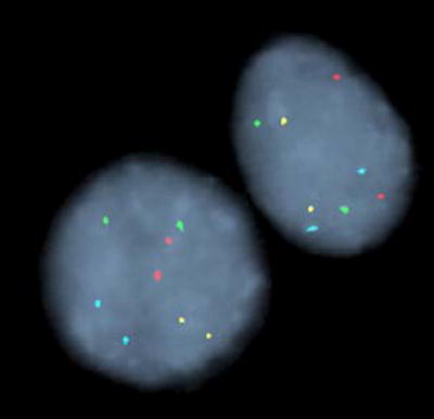
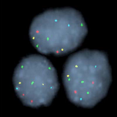
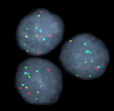
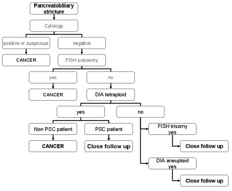
Background & aims: Two advanced cytologic techniques for detecting aneuploidy-digital image analysis (DIA) and fluorescence in situ hybridization (FISH)-have recently been developed to help identify malignant pancreatobiliary strictures. The aim of this study was to assess the clinical utility of cytology, DIA, and FISH for the identification of malignant pancreatobiliary strictures.
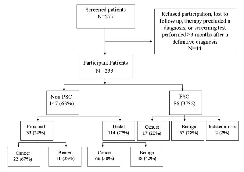
Methods: Brush cytologic specimens from 233 consecutive patients undergoing endoscopic retrograde cholangiopancreatography for pancreatobiliary strictures were examined by all 3 (cytology, DIA, and FISH) techniques. Strictures were stratified as proximal (n = 33) or distal (n = 114) based on whether they occurred above or below the cystic duct, respectively. Strictures in patients with primary sclerosing cholangitis (n = 86) were analyzed separately.
Results: Despite the stratification, the performances of the tests were similar. Conventional cytology has a low sensitivity (4%-20%) but 100% specificity. Because of the high specificity for cytology, we assessed the performance of the other tests when conventional cytology was negative. In this clinical context, FISH had an increased sensitivity (35%-60%) when assessing for chromosomal gains (polysomy) while preserving the specificity of cytology. The sensitivity and specificity of DIA was intermediate as compared with routine cytology and FISH but was additive to FISH values demonstrating only trisomy of chromosome 7 or chromosome 3.
Conclusions: These findings suggest that FISH and DIA increase the sensitivity for the diagnosis of malignant pancreatobiliary tract strictures over that obtained by conventional cytology while maintaining an acceptable specificity.
Figures
References
-
- Kim HJ, Lee KT, Kim SH, Lee JK, Lim JH, Paik SW, Rhee JC. Differential diagnosis of intrahepatic bile duct dilatation without demonstrable mass on ultrasonography or CT: benign versus malignancy. J Gastroenterol Hepatol. 2003;18:1287–92. - PubMed
-
- Rosch T, Hofrichter K, Frimberger E, Meining A, Born P, Weigert N, Allescher HD, Classen M, Barbur M, Schenck U, Werner M. ERCP or EUS for tissue diagnosis of biliary strictures? A prospective comparative study. Gastrointest Endosc. 2004;60:390–6. - PubMed
-
- De Bellis M, Sherman S, Fogel EL, Cramer H, Chappo J, McHenry L, Jr, Watkins JL, Lehman GA. Tissue sampling at ERCP in suspected malignant biliary strictures (Part 1) Gastrointest Endosc. 2002;56:552–61. - PubMed
-
- Nichols JC, Gores GJ, LaRusso NF, Wiesner RH, Nagorney DM, Ritts RE., Jr Diagnostic role of serum CA 19-9 for cholangiocarcinoma in patients with primary sclerosing cholangitis. Mayo Clin Proc. 1993;68:874–9. - PubMed
Publication types
MeSH terms
Substances
Grants and funding
LinkOut - more resources
Full Text Sources
Other Literature Sources
Medical
