. 2006 Oct;131(4):1164-78.
doi: 10.1053/j.gastro.2006.07.019. Epub 2006 Jul 24.

Glial-derived neurotrophic factor modulates enteric neuronal survival and proliferation through neuropeptide Y

Affiliations

Glial-derived neurotrophic factor modulates enteric neuronal survival and proliferation through neuropeptide Y

Mallappa Anitha et al. Gastroenterology. 2006 Oct.

Abstract

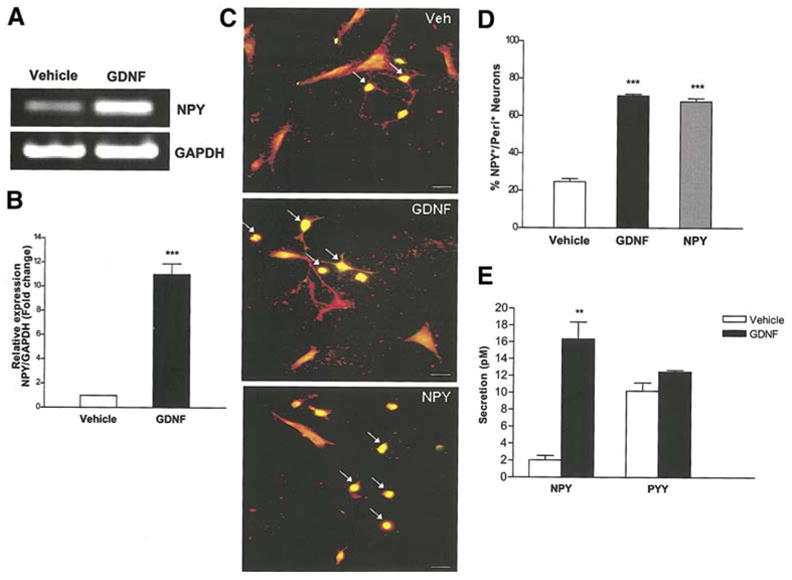
Background & aims: Glial-derived neurotrophic factor (GDNF) promotes the survival and proliferation of enteric neurons. Neuropeptide Y (NPY) is an important peptide regulating gastrointestinal motility. The role of NPY on the survival and proliferation of enteric neurons is not known. We examined the effects of GDNF on the expression and release of NPY from enteric neurons and the role of NPY in promoting enteric neuronal proliferation and survival.

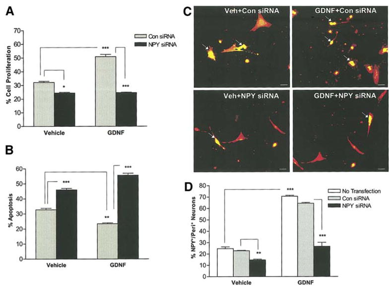
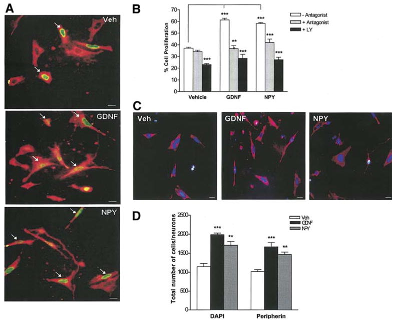
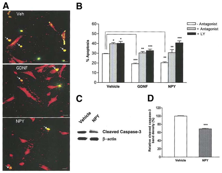
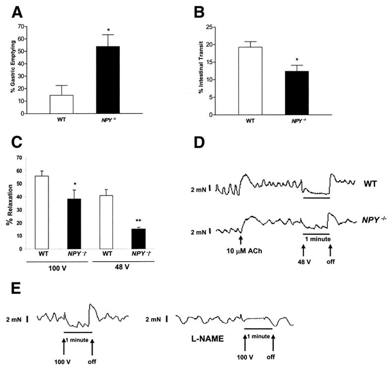
Methods: Studies were performed in primary enteric neuronal cultures and NPY knockout mice (NPY(-/-)). GDNF-induced expression of NPY was assessed by reverse-transcription polymerase chain reaction (RT-PCR), immunocytochemistry, and enzyme-linked immunosorbent assay. Using NPY-siRNA and NPY-Y1 receptor antagonist, we examined the role of NPY in mediating the survival and proliferation effects of GDNF. Gastrointestinal motility was assessed by measuring gastric emptying, intestinal transit, and isometric muscle recording from intestinal muscle strips.

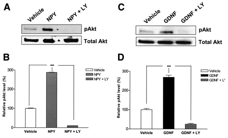
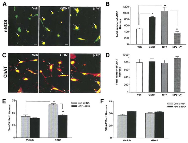
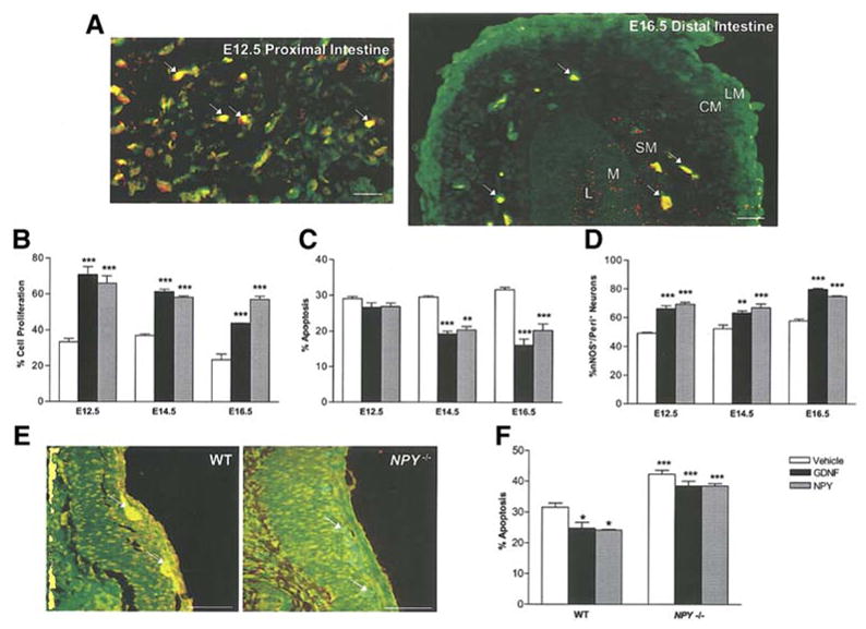
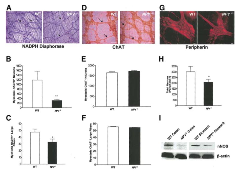
Results: GDNF induced a significant increase in NPY messenger RNA and protein expression in primary enteric neurons and the release of NPY into the culture medium. NPY (1 mumol/L) significantly increased proliferation of neurons and reduced apoptosis. In the presence of NPY-siRNA and NPY-Y1 receptor antagonist or in enteric neurons cultured from NPY(-/-) mice, GDNF-mediated neuronal proliferation and survival was reduced. NPY increased the phosphorylation of Akt, a downstream target of the PI-3-kinase pathway. In NPY(-/-) mice, there were significantly fewer nNOS-containing enteric neurons compared with wild-type (WT) mice. NPY(-/-) mice had accelerated gastric emptying and delayed intestinal transit compared with WT mice.

Conclusions: We demonstrate that NPY acts as an autocrine neurotrophic factor for enteric neurons.


Figures

Figure 1
Figure 1
GDNF increases NPY mRNA and protein expression. (a) RNA was isolated from enteric neurons cultured in the presence or absence of GDNF for 24 h and amplified for GAPDH (loading control) and NPY. The presence of GDNF causes an increase in NPY mRNA. (b) Quantitative real-time PCR showing the relative level of expression of NPY (GDNF vs. vehicle) after normalization to GAPDH expression. Results are mean + S.E., n=9, *** = p<0.001. (c, d) Enteric neurons cultured in the presence of vehicle, GDNF or NPY for 24 h were assessed for peripherin (red)/NPY (green) and the results expressed as % NPY positive neurons. GDNF induced an increase in the number of NPY+ neurons (arrows pointing at yellow neurons). Results are mean + S.E., n=3, *** = p<0.001. Scale bar: 20 μm. (e) Enteric neurons were cultured in the presence or absence of GDNF (48 h) and medium was assayed for NPY and PYY by ELISA. GDNF induced a significant release of NPY but not PYY into the culture medium compared to vehicle. Results are mean + S.E., n=3, ** = p<0.01. P value is with respect to neurons cultured in the presence of vehicle.
Figure 2
Figure 2
GDNF-induced proliferation and survival of enteric neurons is reduced in the presence of NPY siRNA. (a) Enteric neurons in the N2 medium were transfected with control Cyclophilin B or NPY-specific siRNA for 24 h and were subsequently cultured for 24 h in the presence or absence of GDNF. BrdU was added to the medium 24 h prior to the fixation of neurons and the % of BrdU+/peripherin+ neurons was determined to assess the proliferation, n=4. (b) Transfected enteric neurons were assessed for apoptosis using the double-labeling of TUNEL and peripherin, n=3. (c, d) To study the NPY siRNA specificity, transfected enteric neurons were stained for NPY (green)/Peripherin (red), n=3. Results are mean + S.E., *** = p<0.001, ** = p<0.01, * = p<0.05. Scale bar: 20 μm.
Figure 3
Figure 3
GDNF induces proliferation of enteric neurons through NPY. (a, b) Enteric neurons treated with vehicle, NPY or GDNF in the presence or absence of either the NPY-Y1 receptor antagonist (BIBP3226) or the PI-3-kinase inhibitor (LY294002) were maintained in culture for 48 h and fixed. BrdU was added to the medium 24 h prior to fixation. The % of BrdU+ (green)/peripherin+ (red) neurons was determined to assess proliferation. Arrows point to the BrdU+ neurons. Exposure of enteric neurons to NPY (48 h) significantly increased proliferation similar to GDNF. GDNF- and NPY- induced proliferation of enteric neurons was inhibited by both BIBP3226 and LY294002, n=4. Scale bar: 20 μm. (c, d) The total number of cells (DAPI-blue) and neurons (peripherin-red) per well was assessed under the stated culture conditions, n=2. Results are mean + S.E., *** = p<0.001, ** = p<0.01. P value is with respect to the neurons cultured in the absence of antagonist (b) or vehicle (d) in each respective condition.
Figure 4
Figure 4
GDNF suppresses apoptosis of enteric neurons through NPY. (a, b) Enteric neurons treated with vehicle, NPY or GDNF in the presence or absence of BIBP3226 or LY 294002 for 24 h were assessed for apoptosis by the TUNEL (green)/peripherin (red) method. Exposure of enteric neurons to NPY or GDNF significantly reduced apoptosis compared to vehicle. This effect was inhibited by BIBP3226 and LY294002. Arrows show the apoptotic cells with condensed nuclei. Results are mean + S.E., n=3, *** = p<0.001, ** = p<0.01, * = p<0.05. Scale bar: 20 μm. P value is with respect to the neurons cultured in the absence of antagonist in each respective condition. (c, d) Western blot analysis of cleaved caspase-3 in enteric neurons cultured in the presence or absence of NPY for 24 h. β-actin was used as a loading control. There was a significant reduction in the expression of cleaved caspase-3 in NPY treated cells compared to vehicle. Results are ratio + S.E., n=3, *** = p<0.001.
Figure 5
Figure 5
NPY activates the PI-3-kinase pathway. Cell lysates were obtained from enteric neurons treated with vehicle, NPY (a, b) or GDNF (c, d) with or without LY294002 for 30 min and Western blot was performed. GDNF and NPY significantly increased the phosphorylation of Akt compared to vehicle. This was inhibited in the presence of the PI-3-kinase inhibitor LY294002. Results are ratio (pAkt/Akt) ± S.E., n=3, *** = p<0.001.
Figure 6
Figure 6
GDNF and NPY increase nNOS+ enteric neurons but not ChAT+ neurons. Primary enteric neurons were treated with vehicle, NPY, GDNF or NPY+LY294002 for 24 h, fixed and assessed for nNOS (red) and peripherin (green) (a, b) or ChAT (green) and peripherin (red) (c, d), n=4. Scale bar: 20 μm. (e, f) Enteric neurons were transfected with control Cyclophilin B siRNA or NPY specific siRNA for 24 h and then cultured for an additional 24 h in the presence or absence of GDNF, fixed and assessed for nNOS and ChAT, n=3. Results are mean ± S.E., ** = p<0.01, * = p<0.05.
Figure 7
Figure 7
NPY co-localizes with nNOS. Primary enteric neurons were treated with GDNF for 24 h, fixed and assessed for nNOS (red) and peripherin (green) (a), or ChAT (green) and peripherin (red) (b). (c) Percent NPY colocalization with nNOS or ChAT is shown. Results are mean ± S.E., n=3, *** = p<0.001. Scale bar: 20 μm.
Figure 8
Figure 8
Expression and effects of NPY at different developmental stages. (a) NPY is expressed in the proximal intestine of E12.5 rat embryos and extends into the distal intestine in E16.5 rat embryos. NPY is stained with cy3 (red) and peripherin with FITC (green). Arrows show the NPY+ ganglia in E12.5 and E16.5 gut. In the E16.5 intestine the intestinal layers are labeled L (lumen), M (mucosa), SM (submucosal), CM (circular muscle layer), LM (longitudinal muscle layer). Scale bar for (a): 20 μm. (b) The effect of GDNF and NPY on apoptosis and proliferation was assessed in rat embryos at different developmental stages. NPY affects proliferation more in the early stages and apoptosis in the later embryonic stages. (d) nNOS neurons were assessed by immunostaining at different developmental stages. Both GDNF and NPY caused an increase in the number of nNOS+ neurons starting from developmental stage E12.5. (e) Immunohistochemistry for NPY was performed to demonstrate the absence of NPY expression in NPY / mice. The ganglia are stained with peripherin (green) and NPY (red). The overlap of red and green is seen in NPY+ neurons. (f) GDNF did not suppress the apoptosis in enteric neurons cultured from NPY / mice. The effect of GDNF and NPY on apoptosis in enteric neurons obtained from NPY/ mice was assessed by the peripherin/TUNEL method. Results are mean + S.E., *** = p<0.001, ** = p<0.01, * = p<0.05. Scale bar for (e): 100 μm. P value is with respect to the neurons cultured in the presence of vehicle in each respective condition.
Figure 9
Figure 9
NPY/ mice have reduced colonic nNOS+ containing neurons compared to WT mice. (a, b, c) Longitudinal muscle strips along with myenteric plexus from mouse colon were fixed and stained for nNOS+ neurons using the NADPH-diaphorase staining. The nNOS+ neurons and large fibers (arrows) of the myenteric plexus were significantly decreased in the colon of the NPY/ mice compared to WT. (d, e, f) ChAT+ neurons and large fibers (arrows) of the myenteric plexus were similar in the NPY/ and WT colon. Total number of neurons was assessed by peripherin staining (g, h). (i) nNOS expression in WT and NPY / mouse stomach and colon was assessed by Western blot. Scale bar: 100 μm (a, d) and 20 μm (g). Results are mean + S.E., n=6 in each group, ** = p<0.01, * = p<0.05.
Figure 10
Figure 10
Altered motility in NPY/ mice (a) Gastric emptying and intestinal transit was assessed in WT and NPY/ mice. Gastric emptying was significantly higher in NPY/ compared to WT mice. Results are mean + S.E., n=4, * = p<0.05. (b) Intestinal transit was significantly lower in NPY/ compared to WT mice. Results are mean + S.E., n=4, * = p<0.05. (c) Assessment of colonic relaxation with electrical field stimulation in isolated colonic muscle strips containing the myenteric plexus. Transmural electrical field stimulation (100 V/48 V, 20 Hz, 5 msec, 60 s) evoked relaxation of the proximal colon was significantly impaired in NPY/ compared to WT mice. Results are mean + S.E., n=4, * = p<0.05, ** = p<0.01. (d) Representative tracings of electrical field stimulation. The X-axis represents time and the Y-axis represents force in mN. The arrows represent when the electrical stimulation was turned on and off. (e). Effect of L-NAME on electrical field- induced stimulation. The isolated muscle strips from WT mice were pretreated with L-NAME or buffer alone, for 30 min and EFS was performed at 100V, 20 Hz, 5 msec, 60 s. The tracing shows the lack of relaxation in the presence of L-NAME.

References

    1. Heuckeroth RO, Lampe PA, Johnson EM, Milbrandt J. Neurturin and GDNF promote proliferation and survival of enteric neuron and glial progenitors in vitro. Dev Biol. 1998;200:116–29. - PubMed
    1. Gianino S, Grider JR, Cresswell J, Enomoto H, Heuckeroth RO. GDNF availability determines enteric neuron number by controlling precursor proliferation. Development. 2003;130:2187–2198. - PubMed
    1. Young HM, Hearn CJ, Farlie PG, Canty AJ, Thomas PQ, Newgreen DF. GDNF is a chemoattractant for enteric neural cells. Dev Biol. 2001;229:503–16. - PubMed
    1. Natarajan D, Marcos-Gutierrez C, Pachnis V, de Graaff E. Requirement of signalling by receptor tyrosine kinase RET for the directed migration of enteric nervous system progenitor cells during mammalian embryogenesis. Development. 2002;129:5151–60. - PubMed
    1. Barlow A, de Graaff E, Pachnis V. Enteric nervous system progenitors are coordinately controlled by the G protein-coupled receptor EDNRB and the receptor tyrosine kinase RET. Neuron. 2003;40:905–16. - PubMed

Publication types

MeSH terms