

doi: 10.1128/IAI.01429-06.
Epub 2006 Oct 9.
Role of the wbt locus of Francisella tularensis in lipopolysaccharide O-antigen biogenesis and pathogenicity
Affiliations
- PMID: 17030571
- PMCID: PMC1828372
- DOI: 10.1128/IAI.01429-06
Item in Clipboard
Role of the wbt locus of Francisella tularensis in lipopolysaccharide O-antigen biogenesis and pathogenicity
Infect Immun.
2007 Jan.
Abstract
Francisella tularensis is a highly infectious bacterial pathogen, responsible for the zoonotic disease tularemia. We screened a bank of transposon insertion mutants of F. tularensis subsp. holarctica LVS for colony morphology alterations and selected a mutant with a transposon insertion in wbtA, the first gene of the predicted lipopolysaccharide O-antigen gene cluster. Inactivation of wbtA led to the complete loss of O antigen, conferred serum sensitivity, impaired intracellular replication, and severely attenuated virulence in the mouse model. Notably, this mutant afforded protection against a challenge against virulent LVS.
Figures
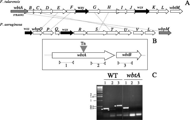
Genetic organization of the F. tularensis wbt locus and transcriptional analysis. (A) Schematic organization of the locus. The arrows indicate approximate sizes and orientations of the different wbt genes. The wzx (determining the cytoplasmic membrane O-antigen transporter), wzy, and wzz (involved in O-antigen polymerization) genes are indicated The LVS wbt locus is shown above the P. aeruginosa O6 wbp locus. Orthologous genes are connected by dotted lines. (B) The transposon insertion site. The filled triangle indicates the transposon (Tn) insertion site in wbtA. The three pairs of primers used for RT-PCR analysis are shown: two pairs in wbtA (to amplify region 1, upstream of the transposon insertion, and region 2, downstream) and one pair in wbtB (region 3). The dotted arrows below indicate the positions of the primers and approximate sizes of the PCR products used in the RT-PCR analysis. (C) RT-PCR. The amplified products were subjected to Tris-acetate-EDTA-agarose gel electrophoresis. RT-PCR was performed on RNA of the LVS wild-type strain (WT) and on RNA of the wbtA mutant. Lane numbers correspond to the regions indicated in panel B. Numbers to the left indicate the molecular mass standards (in kb).
Characteristics of LVS and a wbtA mutant. (A) Morphology on Congo red plates. Overnight cultures were spotted (5 μl) onto solid medium containing Congo red dye (at a final concentration of 100 μg/ml). Morphology alterations were monitored after several (5 to 7) days at 37°C. (B) Growth curves of LVS and mutants in Schaedler K3 broth and in chemically defined medium (2) at 37°C with agitation. (C) Killing of LVS and mutants by nonimmune human serum. Bacteria were incubated in 2% serum; at selected intervals samples were collected, and the number of viable bacteria was determined by plating onto solid medium. (D) Western immunoblotting of whole-cell lysates of LVS and the wbtA mutant. A nitrocellulose membrane was probed with LVS-specific anti-LPS mouse monoclonal T14 antibody at a final dilution of 1:1,000 (left). Numbers to the left correspond to molecular mass markers in kDa. A Coomassie blue-stained gel of the same samples is also shown (right).
Kinetics of intracellular multiplication. Invasiveness of LVS and the wbtA mutant were evaluated in J774 macrophages. Values and error bars represent the means and standard deviations of the number (in log10) of bacteria per well (three wells per assay). The assay was repeated three times (P = 0.028 at 2 h and 3.6 × 10⁻⁵ at 4 h, as determined by a Student's t test).
Virulence of the wbtA mutant. (A) Survival of mice was followed for 9 days after i.p. inoculation with F. tularensis LVS or wbtA mutant bacteria. Groups of five mice were infected with approximately 101, 102, 103, and 104 CFU of LVS or approximately 105, 106, 107, and 108 CFU of the wbtA mutant. (B) Protection of mice against F. tularensis LVS after immunization with the F. tularensis wbtA mutant. Naïve mice or mice immunized 39 days earlier by the i.p. route with 105 CFU of the wbtA mutant were challenged with approximately 104 CFU of LVS, and survival was monitored for 10 days. (C) Growth of F. tularensis LVS and the wbtA mutant in spleen and liver of BALB/c mice. Mice were inoculated by the i.p. route with approximately 104 CFU (five mice per group), and bacterial burdens in spleen and liver were determined 2, 3, 4, and 7 days after infection. †, all mice died. The numbers above the squares indicate the number of mice (in a group of five) in which bacteria were detected (P < 0.001 at all times, as determined by a Student's t test). The arrow indicates the number of CFU used for inoculation.
References
-
- Belanger, M., L. L. Burrows, and J. S. Lam. 1999. Functional analysis of genes responsible for the synthesis of the B-band O antigen of Pseudomonas aeruginosa serotype O6 lipopolysaccharide. Microbiology 145:3505-3521. - PubMed
-
- Cherwonogrodzky, J. W., M. H. Knodel, and M. R. Spence. 1994. Increased encapsulation and virulence of Francisella tularensis live vaccine strain (LVS) by subculturing on synthetic medium. Vaccine 12:773-775. - PubMed
-
- Conlan, J. W., H. Shen, A. Webb, and M. B. Perry. 2002. Mice vaccinated with the O-antigen of Francisella tularensis LVS lipopolysaccharide conjugated to bovine serum albumin develop varying degrees of protective immunity against systemic or aerosol challenge with virulent type A and type B strains of the pathogen. Vaccine 20:3465-3471. - PubMed
-
- Conlan, J. W., E. Vinogradov, M. A. Monteiro, and M. B. Perry. 2003. Mice intradermally inoculated with the intact lipopolysaccharide, but not the lipid A or O-chain, from Francisella tularensis LVS rapidly acquire varying degrees of enhanced resistance against systemic or aerogenic challenge with virulent strains of the pathogen. Microb. Pathog. 34:39-45. - PubMed
Publication types
MeSH terms
Substances
LinkOut - more resources
Full Text Sources
