. 2006 Dec;26(24):9497-507.
doi: 10.1128/MCB.01099-06. Epub 2006 Oct 9.

BRF1 protein turnover and mRNA decay activity are regulated by protein kinase B at the same phosphorylation sites

Affiliations

BRF1 protein turnover and mRNA decay activity are regulated by protein kinase B at the same phosphorylation sites

Don Benjamin et al. Mol Cell Biol. 2006 Dec.

Abstract

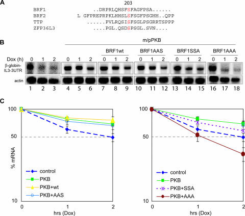
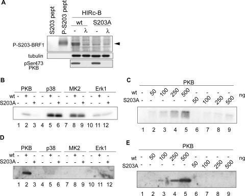
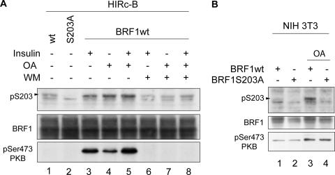
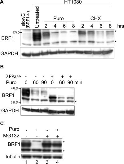
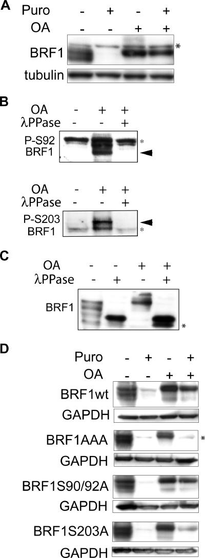
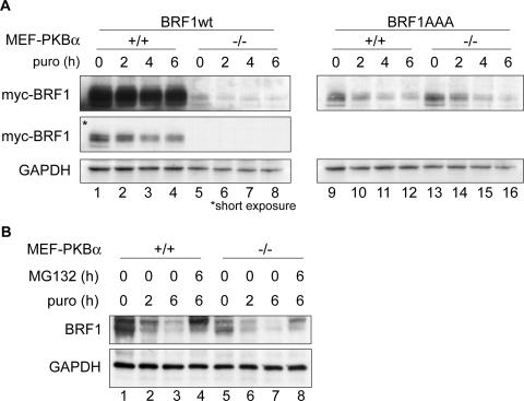
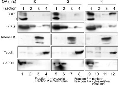
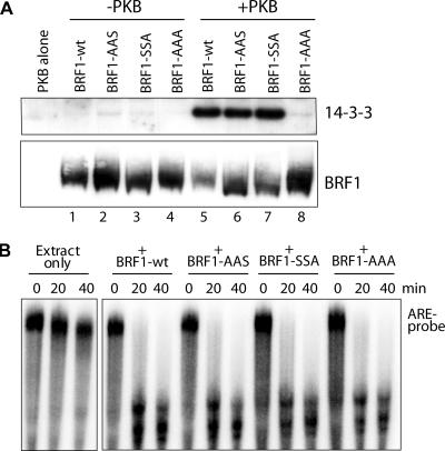
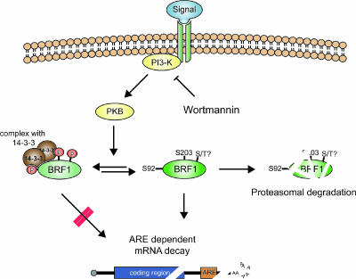
BRF1 posttranscriptionally regulates mRNA levels by targeting ARE-bearing transcripts to the decay machinery. We previously showed that protein kinase B (PKB) phosphorylates BRF1 at Ser92, resulting in binding to 14-3-3 and impairment of mRNA decay activity. Here we identify an additional regulatory site at Ser203 that cooperates in vivo with Ser92. In vitro kinase labeling and wortmannin sensitivity indicate that Ser203 phosphorylation is also performed by PKB. Mutation of both serines to alanine uncouples BRF1 from PKB regulation, leading to constitutive mRNA decay even in the presence of stabilizing signals. BRF1 protein is labile because of proteasomal degradation (half-life, <3 h) but becomes stabilized upon phosphorylation and is less stable in PKBalpha(-/-) cells. Surprisingly, phosphorylation-dependent protein stability is also regulated by Ser92 and Ser203, with parallel phosphorylation required at these sites. Phosphorylation-dependent binding to 14-3-3 is abolished only when both sites are mutated. Cell compartment fractionation experiments support a model in which binding to 14-3-3 sequesters BRF1 through relocalization and prevents it from executing its mRNA decay activity, as well as from proteasomal degradation, thereby maintaining high BRF1 protein levels that are required to reinstate decay upon dissipation of the stabilizing signal.


Figures

FIG. 1.
FIG. 1.
BRF1 activity is cooperatively regulated in vivo at serines 92 and 203. (A) Alignment of the known mammalian ZFP36/Tis11 family members BRF1, BRF2, TTP, and ZFP36L3 reveals a conserved serine at position 203 (BRF1 numbering). The human sequences are shown, except for ZFP36L3, which is mouse specific. The human and mouse BRF1 proteins share perfect homology over the sequence shown. (B) The Tet-β-globin-IL-3 3′ UTR reporter gene was transfected alone (lanes 1 to 3) or in combination with constitutively activated m/pPKB (lanes 4 to 18) and BRF1wt, (lanes 7 to 9), BRF1S90A/S92A (BRF1AAS, lanes 10 to 12), BRF1S203A (BRF1SSA, lanes 13 to 15), or BRF1S90A/S92A/S203A (BRF1AAA, lanes 16 to 18). After 48 h, transcription was stopped by addition of doxycycline (2 μg/ml). Cytoplasmic RNA was isolated at the indicated time points and processed for Northern blotting. Panels for lanes 1 to 3 and lanes 16 to 18 are longer exposures due to lower starting levels of the mRNA reporter. (C) Graphs showing quantification of the reporter transcript signal normalized to the actin control from three independent experiments. Reprobing of the blots for GAPDH and normalization of the reporter probe decay against GAPDH as an independent control gave results similar to those obtained with actin (data not shown).
FIG. 2.
FIG. 2.
PKB phosphorylates BRF1 at serine 203. (A) Characterization of an anti-phospho-Ser203 antibody. HIRc-B cells were transfected with BRF1wt (wt) or the BRF1S203A mutant protein (S203A) and stimulated for 30 min with insulin to activate PKB. Cell lysates were run alongside phosphorylated and unphosphorylated control peptides (pept) to see if the antibody could detect phospho-Ser203 from total cell lysates. Where indicated, the extracts were treated with λ protein phosphatase (30°C, 30 min) before loading. The phosphospecific signal is indicated by the arrowhead. (B) A recombinant BRF1 peptide (aa 143 to 233) containing the wild-type sequence (wt) or the S203A mutation (S203A) was incubated with the activated kinases PKB, p38, MK2, and Erk1 together with radioactive ATP. The samples were resolved, and peptide labeling was visualized by autoradiography. (C) The same conditions for PKB phosphorylation were employed as in panel B with titration of increasing amounts of peptide. (D and E) The preceding experiments were repeated nonradioactively, and the reaction products were subjected to Western blotting with the anti-phospho-Ser203 antibody. Phosphospecific signals were only detected after PKB phosphorylation and were absent from reaction mixtures with the S203A peptide.
FIG. 3.
FIG. 3.
BRF1 is phosphorylated in vivo at serine 203. (A) HIRc-B cells were transfected with BRF1wt or BRF1S203A and stimulated with insulin (20 μg/ml) or 400 nM OA for 30 min at 48 h after transfection. Where indicated, cells were pretreated with WM (200 nM) 15 min before stimulation. Cell lysates were probed with the anti-phospho-Ser203 BRF1 antibody and against anti-phospho-Ser473 PKB to show the correlation between BRF1 Ser203 phosphorylation and PKB activity. (B) NIH 3T3 cells were transfected with BRF1wt or BRF1S203A and stimulated with 400 nM OA for 30 min. The phospho-S203-specific signal is indicated on the left of each panel.
FIG. 4.
FIG. 4.
BRF1 protein is labile. (A) Translation was blocked in HT1080 cells with puromycin (Puro; 50 μg/ml) or 50 μM cycloheximide (CHX), and cells were harvested at the indicated times. Detection with an anti-BRF1 antibody reveals the rapid decay of the protein. The blot was stripped and reprobed against GAPDH as a loading control. Lysate from slowC cells (HT1080 BRF1−/−) was run alongside as a negative control for BRF1. (B) Translation was arrested with puromycin, and cells were harvested at earlier time points. The lysates were also treated with λ phosphatase and run alongside for comparison. (C) Cells were treated for 6 h with puromycin and MG132 (10 μM), a proteasome inhibitor. Stabilization of BRF1 in the presence of MG132 indicates that it is degraded via the proteasome. Asterisks denote nonspecific bands.
FIG. 5.
FIG. 5.
Phosphorylated BRF1 is stable. (A) HT1080 cells were treated with puromycin (Puro; 50 μg/ml) and OA (400 nM) either alone or in combination for 6 h, and lysates were probed with an anti-BRF1 antibody. The persistence of BRF1 in OA-induced cells, even when translation is blocked, indicates that it is stabilized by phosphorylation. Note the upward shift in BRF1 migration characteristic of the hyperphosphorylated form. (B) Probing of lysates from OA-induced cells (400 nM, 3 h) with phosphospecific antibodies directed against phospho-Ser92 and phospho-Ser203 confirms that these sites are phosphorylated in HT1080 cells. Phosphospecific signals (arrowheads) are detected after OA treatment and are sensitive to λ phosphatase (λPPase). (C) Lysates from panel B were probed with the anti-BRF1 antibody. The various BRF1 bands are shifted upward after OA treatment and revert to a single high-mobility band after dephosphorylation by λ phosphatase. (D) A series of BRF1 phospho-null mutants were stably expressed in slowC cells. Translation arrest and OA-induced phosphorylation of BRF1 were performed as described for panel A. Constitutive lability of BRF1AAA protein despite OA-induced hyperphosphorylation suggests that these sites are critical for phosphorylation-dependent stabilization. Unlike the endogenous protein, transfected BRF1 migrates slightly higher than the nonspecific band after OA treatment (compare with panel A) because of the presence of His and Xpress tags. Asterisks indicate nonspecific bands.
FIG. 6.
FIG. 6.
PKB activity affects BRF1 levels. (A) Myc-tagged BRF1wt or the S90/92/203A mutant (BRF1AAA) was transfected into PKBα+/+ and PKBα−/− MEFs. Translation was stopped by the addition of puromycin (puro; 50 μg/ml) for the indicated time periods, and protein levels were determined by Western blotting against the myc tag and GAPDH (loading control). Two different exposures of the Western blot are shown for BRF1wt-transfected cells. (B) Levels of endogenous BRF1 in the MEF cells were determined after translation arrest (puromycin) or addition of the proteasome inhibitor MG132 (10 μM).
FIG. 7.
FIG. 7.
Fractionation of proteins from the major subcellular compartments in control and OA-induced HT1080 cells. Fractionation was performed with 106 cells, and 10 μg of protein was loaded per fraction. Localization of BRF1 and 14-3-3 was determined with the respective antibodies. Reprobing of this blot and a parallel blot made from the same extracts against known marker proteins histone H1 (nuclear), tubulin (cytoskeletal), and GAPDH (cytosolic/membrane) confirms the integrity of the fractionation. The asterisk denotes a nonspecific band.
FIG. 8.
FIG. 8.
Coprecipitation of BRF1-14-3-3 complexes. (A) Recombinant His-BRF1 (40 ng), either unmodified or phosphorylated in vitro by PKBα, was added to 40 μg of slowC cell S100 extract. After binding to nickel beads and washing, the eluted fraction was run on SDS-PAGE and probed against 14-3-3 and BRF1 (as a precipitation control). (B) The various recombinant BRF1 proteins were tested for biological activity by an in vitro RNA decay assay with an ARE RNA probe (39). All proteins could promote rapid decay of the probe, indicating proper conformation and activity.
FIG. 9.
FIG. 9.
A model for coordinated regulation of BRF1 activity and protein stability. In the default state, BRF1 is unphosphorylated at Ser92 and Ser203 and is active in promoting ARE mRNA decay and at the same time is subject to rapid turnover due to proteasomal degradation. A specific mRNA-stabilizing signal results in PKB activation with consequent phosphorylation of these sites. This triggers phosphorylation-dependent BRF1 binding to 14-3-3 and translocation of the complex, which is now unable to promote mRNA decay and cannot be targeted to the proteasome for degradation. An open question is the existence of further phosphoregulatory sites on BRF1, which is extraordinarily serine/threonine rich, that may also regulate its activity by a 14-3-3-independent mechanism.

References

    1. Aitken, A., H. Baxter, T. Dubois, S. Clokie, S. Mackie, K. Mitchell, A. Peden, and E. Zemlickova. 2002. Specificity of 14-3-3 isoform dimer interactions and phosphorylation. Biochem. Soc. Trans. 30:351-360. - PubMed
    1. Andjelković, M., D. R. Alessi, R. Meier, A. Fernandez, N. J. Lamb, M. Frech, P. Cron, P. Cohen, J. M. Lucocq, and B. A. Hemmings. 1997. Role of translocation in the activation and function of protein kinase B. J. Biol. Chem. 272:31515-31524. - PubMed
    1. Baggs, J. E., and C. B. Green. 2003. Nocturnin, a deadenylase in Xenopus laevis retina: a mechanism for posttranscriptional control of circadian-related mRNA. Curr. Biol. 13:189-198. - PubMed
    1. Bakheet, T., M. Frevel, B. R. Williams, W. Greer, and K. S. Khabar. 2001. ARED: human AU-rich element-containing mRNA database reveals an unexpectedly diverse functional repertoire of encoded proteins. Nucleic Acids Res. 29:246-254. - PMC - PubMed
    1. Benjamin, D., M. Colombi, and C. Moroni. 2004. A GFP-based assay for rapid screening of compounds affecting ARE-dependent mRNA turnover. Nucleic Acids Res. 32:e89. - PMC - PubMed

Publication types

MeSH terms