Comparative Study
. 2006 Dec;26(24):9352-63.
doi: 10.1128/MCB.01148-06. Epub 2006 Oct 9.

Neuromedin U receptor 2-deficient mice display differential responses in sensory perception, stress, and feeding

Affiliations
Comparative Study

Neuromedin U receptor 2-deficient mice display differential responses in sensory perception, stress, and feeding

Hongkui Zeng et al. Mol Cell Biol. 2006 Dec.

Abstract

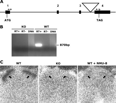
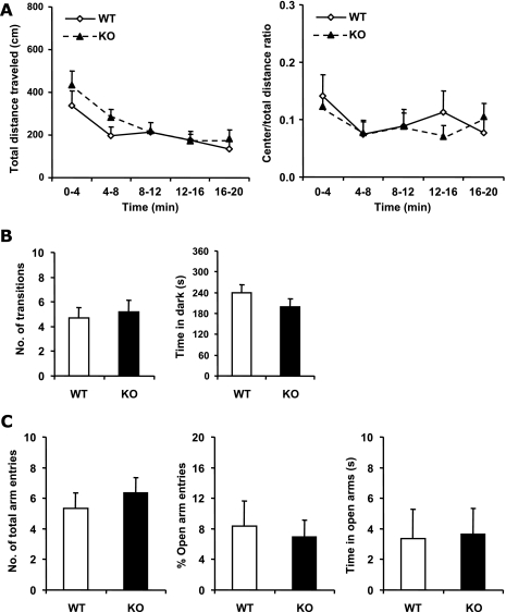
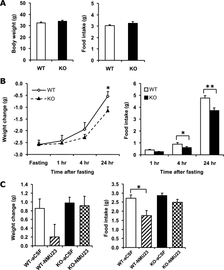
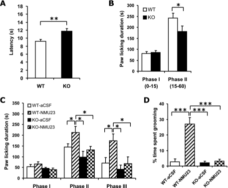
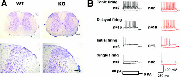
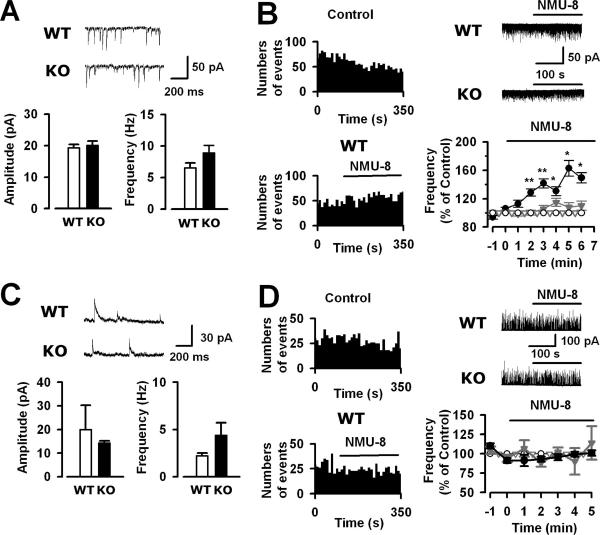
Neuromedin U (NMU) is a highly conserved neuropeptide with a variety of physiological functions mediated by two receptors, peripheral NMUR1 and central nervous system NMUR2. Here we report the generation and phenotypic characterization of mice deficient in the central nervous system receptor NMUR2. We show that behavioral effects, such as suppression of food intake, enhanced pain response, and excessive grooming induced by intracerebroventricular NMU administration were abolished in the NMUR2 knockout (KO) mice, establishing a causal role for NMUR2 in mediating NMU's central effects on these behaviors. In contrast to the NMU peptide-deficient mice, NMUR2 KO mice appeared normal with regard to stress, anxiety, body weight regulation, and food consumption. However, the NMUR2 KO mice showed reduced pain sensitivity in both the hot plate and formalin tests. Furthermore, facilitated excitatory synaptic transmission in spinal dorsal horn neurons, a mechanism by which NMU stimulates pain, did not occur in NMUR2 KO mice. These results provide significant insights into a functional dissection of the differential contribution of peripherally or centrally acting NMU system. They suggest that NMUR2 plays a more significant role in central pain processing than other brain functions including stress/anxiety and regulation of feeding.


Figures

FIG. 1.
FIG. 1.
Generation of NMUR2 KO mice. (A) Schematic diagram of the retroviral insertion in the Nmur2 gene. Numbers 1 to 4 indicate coding exons, with the start codon (ATG) in exon 1 and the stop codon (TAG) in exon 4. The large triangle indicates the viral insertion. Primers a and b are indicated by small arrows. (B) RT-PCR using primers a and b, in which an 870-bp band is expected to be amplified from uninterrupted WT allele. RT+, reverse transcription reaction with the presence of reverse transcriptase. RT−, control reverse transcription reaction without reverse transcriptase. DNA, genomic DNA control in which no PCR band should be amplified because of large intronic regions between the two primers. (C) Receptor binding of 125I-NMU-23 peptide to spinal cord transverse sections from WT and NMUR2 KO mice. Strong binding of 125I-NMU-23 to the outer layers (indicated by arrows) of the spinal dorsal horn is detected in WT but not KO sections. Binding specificity is demonstrated by coincubating 125I-NMU-23 with 1,000-fold excessive unlabeled NMU-8 peptide which results in a complete loss of signal in WT sections. Nonspecific signals in the bones surrounding the spinal cord (as seen partially at the top edges of the micrographs in all three sections) serve as anatomical reference points.
FIG. 2.
FIG. 2.
Comparison of locomotor activity and anxiety between WT and NMUR2 KO mice. (A) Open field activity test. Activity levels are binned every 4 min. Left panel, total distance (in cm) traveled in the open field. Right panel, ratio of distance traveled in the center area versus the total distance traveled. (B) Light-dark box test. Left panel, total number of transitions between the light and dark compartments. Right panel, the time spent in the dark side during the 6-min testing period. (C) Elevated plus maze test. Left panel, total number of entries into both open and closed arms. Middle panel, percentage of entries into the open arms compared with total number of arm entries. Right panel, the time spent in the open arms during the 5-min testing period.
FIG. 3.
FIG. 3.
Comparison of body weights and feeding behavior between WT and NMUR2 KO mice. (A) Body weights (left panel) and 24-h food intake (right panel) of 14- to 17-week-old mice. Each mouse was singly housed for a week before the measuring of body weight and food intake. (B) Fasting and refeeding test. Individually caged mice were fasted for 24 h starting at 1 p.m., with ad lib access to drinking water. Following fasting, mice were weighed and refed. Body weight changes (left panel) relative to prefasting levels and food intake (right panel) were monitored at 1, 4, and 24 h after the start of refeeding. (C) Body weight (left panel) and food intake (right panel) changes 15 h after i.c.v. injection of NMU-23 peptide. To stimulate appetite, mice were fasted for 5 h prior to injection, and NMU-23 (5 nmol in 4 μl aCSF) or vehicle (4 μl aCSF) was administered via i.c.v. injection at 6 p.m. (just before lights out). Food was given back right after injection. At 9 a.m. the following day, food intake and changes in body weight were measured. *, P < 0.05; **, P < 0.01.
FIG. 4.
FIG. 4.
Comparison of nociceptive responses between WT and NMUR2 KO mice. (A) Latencies of hind limb shaking or licking response in hot plate test. (B) Total time spent licking the injected paw in phase I (0 to 15 min after formalin injection) or phase II (15 to 60 min) of the formalin test. (C) Differential effects of NMU-23 in WT and NMUR2 KO mice in the formalin test. Shortly after i.c.v. injection of 3.5 nmol NMU-23 (in 3.5 μl aCSF) or vehicle (3.5 μl aCSF), the mice were placed in the testing container for acclimation for 30 min and then were injected with formalin solution at the right hind paw. The total time spent licking the injected paw in phase I, II, or III (60 to 105 min after formalin injection) is shown. (D) Excessive grooming or BSL behavior after i.c.v. injection of 3.5 nmol NMU-23. This behavior was scored from the videotapes during phase III of the formalin test described for panel C, i.e., 90 to 135 min after the i.c.v. injection, for the percentage of time spent in face-washing, licking or biting all over the body and tail (but excluding the specific licking or biting of the right hind paw injected with formalin solution). *, P < 0.05; **, P < 0.01; ***, P < 0.001.
FIG. 5.
FIG. 5.
Spinal cord morphology and firing patterns of spinal lamina IIo neurons. (A) Cresyl violet staining of lumbar spinal cord transverse sections. Upper panels, lower magnification; scale bar, 200 μm. Lower panels, higher magnification; scale bar, 80 μm. (B) Firing patterns of lamina IIo neurons from WT (traces in black) and NMUR2 KO (traces in red) mice. Twenty-nine WT neurons and 19 KO neurons are similarly categorized into 4 distinct types. No significant differences were revealed between WT and KO neurons in a number of membrane properties, including resting membrane potential (−61.0 ± 1.0 mV, n = 29, versus −63.4 ± 1.9 mV, n = 19), membrane resistance (482.4 ± 33.5 MΩ, n = 29, versus 516.5 ± 48.1 MΩ, n = 19), membrane capacitance (34.2 ± 1.5 pF, n = 29, versus 33.8 ± 2.4 pF, n = 19), and action potential threshold (−34.5 ± 1.7 mV, n = 11, versus −32 ± 1.9 mV, n = 10).
FIG. 6.
FIG. 6.
Effects of NMU-8 on sEPSCs and sIPSCs of spinal lamina IIo neurons in WT and NMUR2 KO mice. (A) Spontaneous EPSCs of lamina IIo neurons: examples (upper traces) and average frequency and amplitude (lower panels). (B) Effect of NMU-8 (10 μM) on sEPSC frequency of lamina IIo neurons. Left panels, time course of WT neurons with control or NMU-8. Upper right panel, traces of WT and KO neurons with NMU-8. Lower right panel, percent change of sEPSC frequency after the application of NMU-8 in WT (black solid circles, n = 20) and KO (gray solid triangles, n = 14) relative to the normalized values in the absence of NMU-8 in WT (black open circles, n = 9) and KO (gray open triangles, n = 5). *, P < 0.05; **, P < 0.01 between WT NMU-8 and control curve. (C) Spontaneous IPSCs of lamina IIo neurons: examples (upper traces) and average frequency and amplitude (lower panels). (D) Effect of NMU-8 (10 μM) on sIPSC frequency of lamina IIo neurons. Left panels, time course of WT neurons with control or NMU-8. Upper right panel, traces of WT and KO neurons with NMU-8. Lower right panel, percent change of sIPSC frequency after the application of NMU-8 in WT (black solid circles, n = 5) and KO (gray solid triangles, n = 4) relative to the normalized values in the absence of NMU-8 in WT (black open circles, n = 5) and KO (gray open triangles, n = 4).

References

    1. Amit, Z., and Z. H. Galina. 1986. Stress-induced analgesia: adaptive pain suppression. Physiol. Rev. 66:1091-1120. - PubMed
    1. Arancio, O., E. R. Kandel, and R. D. Hawkins. 1995. Activity-dependent long-term enhancement of transmitter release by presynaptic 3′,5′-cyclic GMP in cultured hippocampal neurons. Nature 376:74-80. - PubMed
    1. Brighton, P. J., P. G. Szekeres, and G. B. Willars. 2004. Neuromedin U and its receptors: structure, function, and physiological roles. Pharmacol. Rev. 56:231-248. - PubMed
    1. Cao, C. Q., X. H. Yu, A. Dray, A. Filosa, and M. N. Perkins. 2003. A pro-nociceptive role of neuromedin U in adult mice. Pain 104:609-616. - PubMed
    1. Craig, A. D. 1996. An ascending general homeostatic afferent pathway originating in lamina I. Prog. Brain Res. 107:225-242. - PubMed

Publication types