. 2006 Oct 17;103(42):15491-6.
doi: 10.1073/pnas.0607538103. Epub 2006 Oct 9.

Angiopoietin-2 functions as an autocrine protective factor in stressed endothelial cells

Affiliations

Angiopoietin-2 functions as an autocrine protective factor in stressed endothelial cells

Christopher Daly et al. Proc Natl Acad Sci U S A. .

Abstract

Angiopoietin (Ang)-2, a context-dependent agonist/antagonist for the vascular-specific Tie2 receptor, is highly expressed by endothelial cells at sites of normal and pathologic angiogenesis. One prevailing model suggests that in these settings, Ang-2 acts as an autocrine Tie2 blocker, inhibiting the stabilizing influence of the Tie2 activator Ang-1, thereby promoting vascular remodeling. However, the effects of endogenous Ang-2 on cells that are actively producing it have not been studied in detail. Here, we demonstrate that Ang-2 expression is rapidly induced in endothelial cells by the transcription factor FOXO1 after inhibition of the phosphatidylinositol 3-kinase/Akt pathway. We employ RNAi and blocking antibodies to show that in this setting, Ang-2 unexpectedly functions as a Tie2 agonist, bolstering Akt activity so as to provide negative feedback on FOXO1-regulated transcription and apoptosis. In addition, we show that Ang-2, like Ang-1, activates Tie2/Akt signaling in vivo, thereby inhibiting the expression of FOXO1 target genes. Consistent with a role for Ang-2 as a Tie2 activator, we demonstrate that Ang-2 inhibits vascular leak. Our data suggests a model in which Ang-2 expression is induced in stressed endothelial cells, where it acts as an autocrine Tie2 agonist and protective factor.


Conflict of interest statement

Conflict of interest statement: All of the authors except one (D.M.M.) are employees of, and own stock or stock options in, Regeneron Pharmaceuticals.

Figures

Fig. 1.
Fig. 1.
Inhibition of phosphatidylinositol 3-kinase/Akt signaling results in a rapid FOXO1-dependent increase in Ang-2 expression. (A) HUVECs were either untreated or treated with 0.25 μM WM for 60 min. Cell lysates were prepared and subjected to Western blot with antibodies against phospho-Ser-473 Akt (p-Akt) and Akt. (B) HUVECs were either untreated or treated with 0.25 μM WM for 15, 30, or 60 min. Cytoplasmic (C) and nuclear (N) fractions were prepared and subjected to Western blot with antibodies against FOXO1, nuclear lamin, and ERK. (C) HUVECs were infected with viruses encoding either control siRNA or FOXO1-specific siRNA. At 24 and 48 h after infection, cell lysates were prepared and subjected to Western blot with antibodies against FOXO1 and ERK. (D) HUVECs were infected with viruses encoding either control siRNA or FOXO1-specific siRNA. At 30 h after infection, cells were either untreated (−) or treated with WM for 2 h. Total RNA was isolated and the levels of Ang-2 RNA (normalized to actin levels) were determined by real-time PCR. The data are expressed as relative Ang-2 level vs. untreated cells in the presence of control siRNA (assigned a value of 1.0). Bars represent mean and SD; n = 3.
Fig. 2.
Fig. 2.
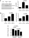
FOXO1 induces Ang-2 expression and promotes Tie2 phosphorylation. (A) HUVECs were infected with adenoviruses encoding either GFP or FOXO1-TM for 20 h. Cell lysates were prepared and subjected to Western blot with antibodies against Ang-2 and ERK. (B) Tissue culture medium was collected from HUVECs expressing GFP or FOXO1-TM for 20 h, and Ang-2 levels were determined by ELISA. Bars represent mean and SD; n = 3. (C and D) HUVECs (C) or BLMVECs (D) were infected with adenoviruses encoding either GFP or FOXO1-TM for 20 h. Tie2 was immunoprecipitated and subjected to Western blot with antibodies against phosphotyrosine (p-Tie2) and Tie2. The graphs show the change in p-Tie2/Tie2 ratio induced by FOXO1-TM. Bars represent mean and SD; n = 3.
Fig. 3.
Fig. 3.
FOXO1-induced Tie2 phosphorylation depends on endogenous Ang-2. (A) HUVECs were infected with adenoviruses encoding either control siRNA or Ang-2-specific siRNA. At 48 h after infection, cell lysates were prepared and subjected to Western blot with antibodies against Ang-2 and ERK. (B) Ang-2 protein levels were assessed by Western blot at 30 or 48 h (48 h data are shown in A) after expression of control or Ang-2 siRNA. The data are expressed as relative Ang-2 protein level vs. control siRNA treatment (assigned a value of 1.0). Bars represent mean and SD; n = 3. (C) HUVECs were infected with adenoviruses encoding either control or Ang-2-specific siRNA. Forty-eight hours later, cells were infected with viruses encoding either GFP or FOXO1-TM for 20 h. Tie2 was immunoprecipitated and subjected to Western blot with antibodies against phosphotyrosine (p-Tie2) and Tie2. The relative p-Tie2/Tie2 ratios are shown in the graph. Bars represent mean and SD; n = 3. In addition, aliquots of the whole-cell lysates were subjected to Western blot with antibodies against Ang-2 and FOXO1 (myc tag antibody). (D) HUVECs were infected with adenoviruses encoding either GFP or FOXO1-TM in the presence of control antibody or Ang-2 antibody. At 20 h after infection, Tie2 was immunoprecipitated and subjected to Western blot with antibodies against phosphotyrosine (p-Tie2) and Tie2. The relative p-Tie2/Tie2 ratios are shown in the graph. Bars represent mean and SD; n = 3. (E) HUVECs were infected with adenoviruses encoding either GFP or FOXO1-TM in the presence of control peptide-Fc fusion protein or Ang-2-specific peptide-Fc fusion protein. At 20 h after infection, Tie2 was immunoprecipitated and subjected to Western blot with antibodies against phosphotyrosine (p-Tie2) and Tie2.
Fig. 4.
Fig. 4.
FOXO1-induced Ang-2 activates Akt and inhibits FOXO1-dependent transcription and apoptosis. (A) BLMVECs were infected with adenoviruses encoding GFP or native FOXO1 in the presence of either control antibody or Ang-2 antibody. At 20 h after infection, cell lysates were prepared and subjected to Western blot with antibodies against phospho-Ser-473 Akt (p-Akt) or Akt. The graph shows the relative p-Akt/Akt ratios. Bars represent mean and SD; n = 3. (B) BLMVECs were infected with adenoviruses encoding GFP or native FOXO1 in the presence of either control antibody or Ang-2 antibody. At 20 h after infection, RNA was isolated, and the levels of Ang-2 and decorin RNA (normalized to GAPDH levels) were determined by real-time PCR. The data are expressed as relative Ang-2 or decorin level vs. GFP/control Ab samples (assigned a value of 1.0). The bars represent mean and SD; n = 3. (C) BLMVECs were infected with adenoviruses encoding GFP or native FOXO1 in the presence of either control antibody or Ang-2 antibody. At 24 h after the start of infection, cells were subjected to serum starvation for an additional 72 h in the presence of control or Ang-2 antibody, and the number of viable cells was determined by MTS assay. The graph shows the number of living cells relative to the GFP/control Ab samples (assigned a value of 1.0). The bars represent mean and SD; n = 3.
Fig. 5.
Fig. 5.
Ang-2 activates Tie2/Akt signaling, inhibits expression of FOXO1 target genes, and inhibits vascular leak in vivo. (A) Mice were injected i.v. with 200 μg of human Fc, Ang-1 (A1), or Ang-2 (A2). Heart extracts were prepared at 15, 30, or 60 min after injection. Tie2 was immunoprecipitated and subjected to Western blot with antibodies against phosphotyrosine (p-Tie2) and Tie2. (B) Mice were injected intravenously with 200 μg of human Fc, Ang-1 (A1), or Ang-2 (A2). At 15 or 30 min after injection, heart extracts were prepared and subjected to Western blot with antibodies against phospho-Ser-473 Akt (p-Akt) and Akt. (C) Mice were injected i.v. with adenoviruses (109 pfu per animal) encoding Fc, Ang-1 (A1), or Ang-2 (A2). At 24 h after infection, RNA was isolated from heart tissue and the levels of Ang-2 and ESM-1 RNA (normalized to actin levels) were determined by real-time PCR. The data are expressed as relative Ang-2 or ESM-1 level vs. Fc controls (assigned a value of 1.0). The bars represent mean and SD; n = 3. The Ang-1- and Ang-2-mediated changes in expression of both target genes were statistically significant at P < 0.01. (D) Mice were either uninfected or infected with adenoviruses (109 pfu per animal) encoding Ang-1 (A1) or Ang-2 (A2). At 3 days after infection, mice were injected with Evans blue dye, and ears were either untreated or treated with mustard oil. Extravasated Evans blue was quantitated spectrophotometrically. The graph shows the mean and SE. The effects of both Ang-1 and Ang-2 on mustard oil-induced vascular leak were statistically significant, P < 0.01.

References

    1. Sato TN, Tozawa Y, Deutsch U, Wolburg-Buchholz K, Fujiwara Y, Gendron-Maguire M, Gridley T, Wolburg H, Risau W, Qin Y. Nature. 1995;376:70–74. - PubMed
    1. Davis S, Aldrich TH, Jones PF, Acheson A, Compton DL, Jain V, Ryan TE, Bruno J, Radziejewski C, Maisonpierre PC, Yancopoulos GD. Cell. 1996;87:1161–1169. - PubMed
    1. Suri C, Jones PF, Patan S, Bartunkova S, Maisonpierre PC, Davis S, Sato TN, Yancopoulos GD. Cell. 1996;87:1171–1180. - PubMed
    1. Davis S, Papadopoulos N, Aldrich TH, Maisonpierre PC, Huang T, Kovac L, Xu A, Leidich R, Radziejewska E, Rafique A, et al. Nat Struct Biol. 2003;10:38–44. - PubMed
    1. Maisonpierre PC, Suri C, Jones PF, Bartunkova S, Wiegand SJ, Radziejewski C, Compton D, McClain J, Aldrich TH, Papadopoulos N, et al. Science. 1997;277:55–60. - PubMed

MeSH terms