

An ex vivo study of the biological properties of porcine aortic valves in response to circumferential cyclic stretch
- PMID: 17031600
- PMCID: PMC1705516
- DOI: 10.1007/s10439-006-9167-8
An ex vivo study of the biological properties of porcine aortic valves in response to circumferential cyclic stretch
Abstract
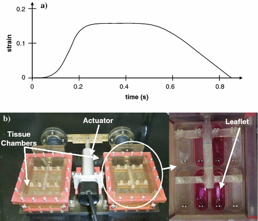
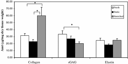
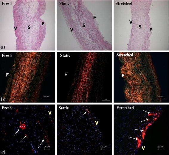
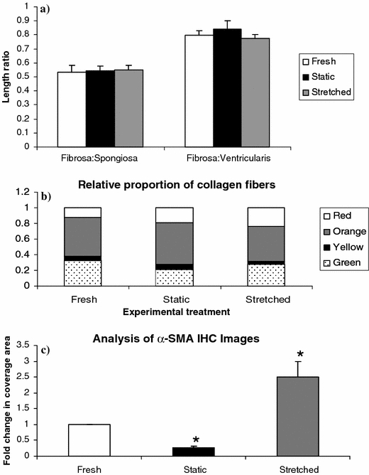
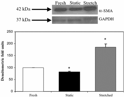
Normal physiological mechanical forces cause constant tissue renewal in aortic valve leaflets (AVL) while altered mechanical forces incite changes in their structural and biological properties. The current study aims at characterizing the remodeling properties of AVL subjected to cyclic circumferential stretch in a sterile ex vivo bioreactor. The leaflets cultured were stretched at a maximum rate of 300%s(-1) corresponding to a 15% strain for 48 h. Collagen, sulfated glycosaminoglycan (sGAG), and elastin contents of the stretched, fresh, and statically incubated leaflets were measured. Cusp morphology and cell phenotype were also examined. AVLs exposed to cyclic stretch showed a significant increase in collagen content (p < 0.05) when compared to fresh and statically incubated AVLs. sGAG content was significantly reduced in the stretched AVLs (p < 0.05) when compared to the fresh leaflets and was comparable between stretched and statically incubated AVLs. There was no statistically significant change in elastin content in all the three groups of AVLs (p > 0.05). Native aortic valve morphology was well preserved in stretched leaflets. Immunohistochemistry and immunoblotting studies showed an increased expression of alpha-smooth muscle actin (alpha-SMA) in stretched leaflets while alpha-SMA expression was reduced in statically incubated AVLs when compared to the fresh leaflets. To conclude, circumferential cyclic stretch altered the extracellular matrix remodeling activity of valvular cells, and consequently the extracellular matrix composition of the AVLs. Most interestingly, the contractile and fibrotic phenotypic expression of valve interstitial cells was enhanced. These results show that circumferential cyclic stretch is a possible mediator for AVL remodeling activity.
Figures
References
-
- {'text': '', 'ref_index': 1, 'ids': [{'type': 'DOI', 'value': '10.1016/S0735-1097(01)01422-X', 'is_inner': False, 'url': 'https://doi.org/10.1016/s0735-1097(01)01422-x'}, {'type': 'PubMed', 'value': '11527641', 'is_inner': True, 'url': 'https://pubmed.ncbi.nlm.nih.gov/11527641/'}]}
- Agmon Y., et al. (2001) Aortic valve sclerosis and aortic atherosclerosis: different manifestations of the same disease? Insights from a population-based study J. Am. Coll. Cardiol. 38(3): 827–834 - PubMed
-
- {'text': '', 'ref_index': 1, 'ids': [{'type': 'DOI', 'value': '10.1161/CIRCULATIONAHA.105.591768', 'is_inner': False, 'url': 'https://doi.org/10.1161/circulationaha.105.591768'}, {'type': 'PubMed', 'value': '16534030', 'is_inner': True, 'url': 'https://pubmed.ncbi.nlm.nih.gov/16534030/'}]}
- Aikawa E., et al. (2006) Human semilunar cardiac valve remodeling by activated cells from fetus to adult: implications for postnatal adaptation, pathology, and tissue engineering Circulation 113(10): 1344–1352 - PubMed
-
- {'text': '', 'ref_index': 1, 'ids': [{'type': 'DOI', 'value': '10.1161/01.RES.0000141146.95728.da', 'is_inner': False, 'url': 'https://doi.org/10.1161/01.res.0000141146.95728.da'}, {'type': 'PMC', 'value': 'PMC2810618', 'is_inner': False, 'url': 'https://pmc.ncbi.nlm.nih.gov/articles/PMC2810618/'}, {'type': 'PubMed', 'value': '15345668', 'is_inner': True, 'url': 'https://pubmed.ncbi.nlm.nih.gov/15345668/'}]}
- Armstrong E. J., Bischoff J., (2004) Heart valve development: endothelial cell signaling and differentiation Circ. Res. 95(5): 459–470 - PMC - PubMed
-
- {'text': '', 'ref_index': 1, 'ids': [{'type': 'DOI', 'value': '10.1161/01.ATV.0000130462.50769.5a', 'is_inner': False, 'url': 'https://doi.org/10.1161/01.atv.0000130462.50769.5a'}, {'type': 'PubMed', 'value': '15117733', 'is_inner': True, 'url': 'https://pubmed.ncbi.nlm.nih.gov/15117733/'}]}
- Butcher J. T., et al. (2004) Unique morphology and focal adhesion development of valvular endothelial cells in static and fluid flow environments Arterioscler. Thromb. Vasc. Biol. 24(8): 1429–1434 - PubMed
-
- {'text': '', 'ref_index': 1, 'ids': [{'type': 'PubMed', 'value': '15222296', 'is_inner': True, 'url': 'https://pubmed.ncbi.nlm.nih.gov/15222296/'}]}
- Butcher J. T., Nerem R. M., (2004) Porcine aortic valve interstitial cells in three-dimensional culture: comparison of phenotype with aortic smooth muscle cells J. Heart. Valve Dis. 13(3): 478–485; (discussion 485–486) - PubMed
Publication types
MeSH terms
Substances
LinkOut - more resources
Full Text Sources
