. 2006 Nov;210(3):288-97.
doi: 10.1002/path.2067.

Expression of elevated levels of pro-inflammatory cytokines in SARS-CoV-infected ACE2+ cells in SARS patients: relation to the acute lung injury and pathogenesis of SARS

Affiliations

Expression of elevated levels of pro-inflammatory cytokines in SARS-CoV-infected ACE2+ cells in SARS patients: relation to the acute lung injury and pathogenesis of SARS

L He et al. J Pathol. 2006 Nov.

Abstract

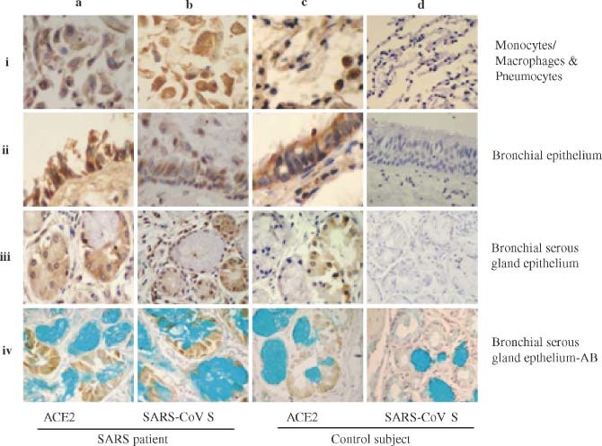
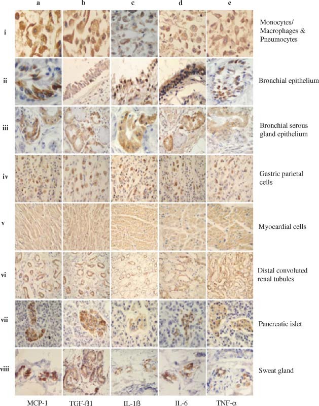
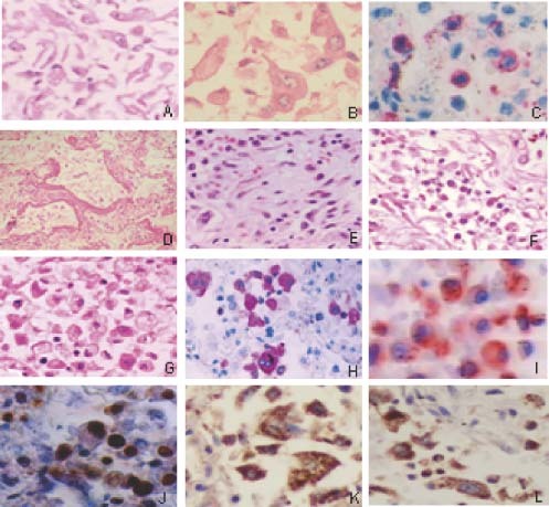
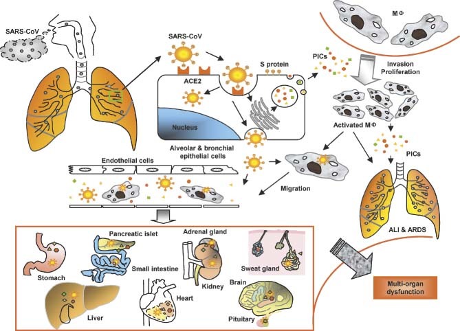
The authors have previously shown that acute lung injury (ALI) produces a wide spectrum of pathological processes in patients who die of severe acute respiratory syndrome (SARS) and that the SARS coronavirus (SARS-CoV) nucleoprotein is detectable in the lungs, and other organs and tissues, in these patients. In the present study, immunohistochemistry (IHC) and in situ hybridization (ISH) assays were used to analyse the expression of angiotensin-converting enzyme 2 (ACE2), SARS-CoV spike (S) protein, and some pro-inflammatory cytokines (PICs) including MCP-1, TGF-beta1, TNF-alpha, IL-1beta, and IL-6 in autopsy tissues from four patients who died of SARS. SARS-CoV S protein and its RNA were only detected in ACE2+ cells in the lungs and other organs, indicating that ACE2-expressing cells are the primary targets for SARS-CoV infection in vivo in humans. High levels of PICs were expressed in the SARS-CoV-infected ACE2+ cells, but not in the uninfected cells. These results suggest that cells infected by SARS-CoV produce elevated levels of PICs which may cause immuno-mediated damage to the lungs and other organs, resulting in ALI and, subsequently, multi-organ dysfunction. Therefore application of PIC antagonists may reduce the severity and mortality of SARS.


Figures

Figure 1
Figure 1
Detection of ACE2 and SARS‐CoV S protein in the lung and bronchial tissues by IHC. The MAbs against human ACE2 and the SARS‐CoV S protein were used to detect the expression of ACE2 (a–i to a–iv and c–i to c–iv) and SARS‐CoV S protein (b–i to b–iv and d–i to d–iv) in monocytes/macrophages and pneumocytes (a–i to d–i), bronchial epithelial cells (a–ii to d–ii), bronchial serous gland epithelium (a–iii to d–iii), and bronchial serous gland epithelium‐AB (a–iv to d–iv) in the SARS patients (a–i to b–iv) and control subject (c–i to d–iv). All sections were counter‐stained with haematoxylin, except the sections of bronchial serous gland epithelium‐AB, which were counter‐stained with haematoxylin and Alcian blue
Figure 2
Figure 2
Detection of ACE2 and SARS‐CoV S protein in other organs and tissues by IHC. The MAbs against human ACE2 and the SARS‐CoV S protein were used to detect the expression of ACE2 (a–i to a–v and c–i to c–v) and SARS‐CoV S protein (b–i to b–v and d–i to d–v) in myocardial cells (a–i to d–i), gastric parietal cells (a–ii to d–ii), distal convoluted renal tubules (a–iii to d–iii), pancreatic islet (a–iv to d–iv), and sweat gland (a–v to d–v) in the SARS patients (a–i to b–v) and control subject (c–i to d–v). All sections were counter‐stained with haematoxylin
Figure 3
Figure 3
Detection of pro‐inflammatory cytokines in the lungs and other organs and tissues by IHC. The MAbs against human MCP‐1, TGF‐β1, IL‐1β, IL‐6, and TNF‐α, respectively, were used to detect the expression of MCP‐1 (a–i to a–viii), TGF‐β1 (b–i to b–viii), IL‐1β (c–i to c–viii), IL‐6 (d–i to d–viii), and TNF‐α (e–i to e–viii) in monocytes/macrophages and pneumocytes (a–i to e–i), bronchial epithelial cells (a–ii to e–ii), bronchial serous gland epithelium (a–iii to e–iii), gastric parietal cells (a–iv to e–iv), myocardial cells (a–v to e–v), distal convoluted renal tubules (a–vi to e–vi), pancreatic islet (a–vii to e–vii), and sweat gland cells (a–viii to e–viii) in the SARS patients. All sections were counter‐stained with haematoxylin
Figure 4
Figure 4
Pathological changes in the lungs of SARS patients. (A) Alveoli filled with desquamated epithelial cells (H&E); (B) mononuclear and multinucleate epithelial giant cells in alveoli (H&E); (C) T‐lymphocyte infiltrates (CD45RO staining); (D) formation of hyaline membranes (H&E); (E) organization and fibrinosis of alveolar exudates; (F) exudation of monocytes/macrophages and lymphocytes in pulmonary interstitial tissues (H&E); (G) exudation of monocytes/macrophages and lymphocyte in alveoli (H&E); (H) obvious proliferation of macrophages in the alveoli cavities (Mac387 staining positive); (I) active macrophages in the alveoli (CD25 staining positive); and apoptosis in the pneumocytes as shown by Klenow‐FragEL (J), Fas (K), and FasL (L) staining
Figure 5
Figure 5
A model for the immunopathogenesis of SARS. SARS‐CoV in droplets enters into the lung, where the virus binds via its S protein to ACE2 on the alveolar or bronchial epithelial cells. The virus replicates in these cells, from which new virions are released into the blood. The infected cells under the stimulation of SARS‐CoV and some uninfected cells induced by viral antigens or PIC‐regulatory factors produce high levels of PICs to mediate inflammatory responses for combating the virus. However, these PICs also damage the host cells. Some of the PICs, eg monocyte chemoattractant protein‐1 (MCP‐1), attract monocytes in blood to migrate to the alveolar cavities, where the monocytes are stimulated by other PICs to become proliferative and/or activated macrophages (MΦ). The activated macrophages can produce more PICs and may transmit SARS‐CoV to other sites. Some of the PICs, including TGF‐β1 and TNF‐α, may induce apoptotic death of the epithelial cells, pneumocytes, and lymphocytes, or mediate pulmonary fibronosis, resulting in ALI and ARDS. The cell‐free and MΦ‐associated SARS‐CoV in the blood can be transmitted from the lung to other organs to infect the ACE2‐expressing cells in the local sites. More PICs are produced and the level of PICs in the blood is rapidly elevated, leading to multi‐organ dysfunction. MΦ = macrophages; ALI = acute lung injury; PICs = pro‐inflammatory cytokines

References

    1. Ksiazek TG, Erdman D, Goldsmith CS, Zaki SR, Peret T, Emery S, et al. A novel coronavirus associated with severe acute respiratory syndrome. N Engl J Med 2003; 348: 1953–1966. - PubMed
    1. Drosten C, Gunther S, Preiser W, Van Der WS, Brodt HR, Becker S, et al. Identification of a novel coronavirus in patients with severe acute respiratory syndrome. N Engl J Med 2003; 348: 1967–1976. - PubMed
    1. Peiris JS, Lai ST, Poon LL, Guan Y, Yam LY, Lim W, et al. Coronavirus as a possible cause of severe acute respiratory syndrome. Lancet 2003; 361: 1319–1325. - PMC - PubMed
    1. Marra MA, Jones SJM, Astell CR, Holt RA, Brooks‐Wilson A, Butterfield YSN, et al. The genome sequence of the SARS‐associated coronavirus. Science 2003; 300: 1399–1404. - PubMed
    1. Rota PA, Oberste MS, Monroe SS, Nix WA, Campagnoli R, Icenogle JP, et al. Characterization of a novel coronavirus associated with severe acute respiratory syndrome. Science 2003; 300: 1394–1399. - PubMed

Publication types

MeSH terms