Not all age-related white matter hyperintensities are the same: a magnetization transfer imaging study
- PMID: 17032876
- PMCID: PMC7977888
Not all age-related white matter hyperintensities are the same: a magnetization transfer imaging study
Abstract
Purpose: Our aim was to assess whether presumed histologic heterogeneity of age-related white matter hyperintensities (WMH) is reflected in quantitative magnetization transfer imaging measures.
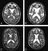
Materials and methods: From a group of patients participating in a double-blind placebo-controlled multicenter study on the effect of pravastatin (PROSPER), we selected 56 subjects with WMH. WMH were classified as periventricular WMH (PVWMH) and deep WMH (DWMH). PVWMH were subclassified as irregular or smooth, depending on the aspect of their border. Signal intensity of WMH on T1-weighted images was scored as iso- or hypointense. The mean magnetization transfer ratio (MTR) value of different types of WMH was assessed and compared. As a control group, we selected 19 subjects with no or limited WMH.
Results: Mean (SE) MTR of PVWMH (frontal, 31.2% [0.2%]; occipital, 32.2% [0.2%]) was lower than that of DWMH (33.7% [0.5%]). The mean MTR of frontal PVWMH (31.2% [0.2%]) was lower than that of occipital PVWMH (32.2% [0.2%]). Compared with occipital PVWMH, frontal PVWMH more often had a smooth lining (72% frontal versus 8% occipital) and an area with low signal intensity on T1-weighted images (76% frontal versus 35% occipital). MTR did not differ between smooth (31.1% [0.3%]) and irregular (31.6% [0.5%]) PVWMH.
Conclusion: Age-related WMH are heterogeneous, despite their similar appearance on T2-weighted images. By taking into account heterogeneity of age-related WMH, both in terms of etiology and in terms of severity of tissue destruction, one may obtain better understanding on the causes and consequences of these lesions.
Figures


References
-
- Meyer JS, Kawamura J, Terayama Y. White matter lesions in the elderly. J Neurol Sci 1992;110:1–7 - PubMed
-
- de Leeuw FE, de Groot JC, Oudkerk M, et al. Aortic atherosclerosis at middle age predicts cerebral white matter lesions in the elderly. Stroke 2000;31:425–29 - PubMed
-
- Longstreth WT Jr, Manolio TA, Arnold A, et al. Clinical correlates of white matter findings on cranial magnetic resonance imaging of 3301 elderly people: The Cardiovascular Health Study. Stroke 1996;27:1274–82. Comment in: Stroke 1996;27:1269–73, Stroke 1996;27:2338 - PubMed
-
- Breteler MM, van Amerongen NM, van Swieten JC, et al. Cognitive correlates of ventricular enlargement and cerebral white matter lesions on magnetic resonance imaging: The Rotterdam Study. Stroke 1994;25:1109–15 - PubMed
Publication types
MeSH terms
Substances
LinkOut - more resources
Full Text Sources
Medical
Research Materials
