

Improved delineation of glioma margins and regions of infiltration with the use of diffusion tensor imaging: an image-guided biopsy study
- PMID: 17032877
- PMCID: PMC7977915
Improved delineation of glioma margins and regions of infiltration with the use of diffusion tensor imaging: an image-guided biopsy study
Abstract
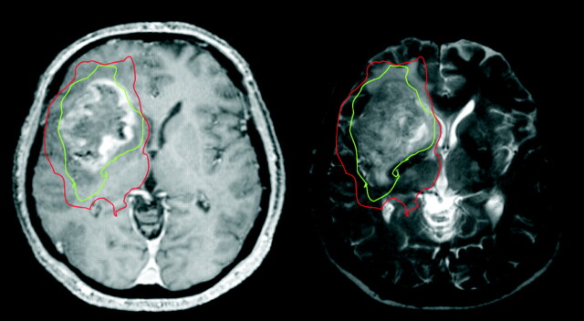
Background and purpose: The efficacy of radiation therapy, the mainstay of treatment for malignant gliomas, is limited by our inability to accurately determine tumor margins. As a result, despite recent advances, the prognosis remains appalling. Because gliomas preferentially infiltrate along white matter tracks, methods that show white matter disruption should improve this delineation. In this study, results of histologic examination from samples obtained from image-guided brain biopsies were correlated with diffusion tensor images.
Methods: Twenty patients requiring image-guided biopsies for presumed gliomas were imaged preoperatively. Patients underwent image-guided biopsies with multiple biopsies taken along a single track that went into normal-appearing brain. Regions of interest were determined from the sites of the biopsies, and diffusion tensor imaging findings were compared with glioma histology.
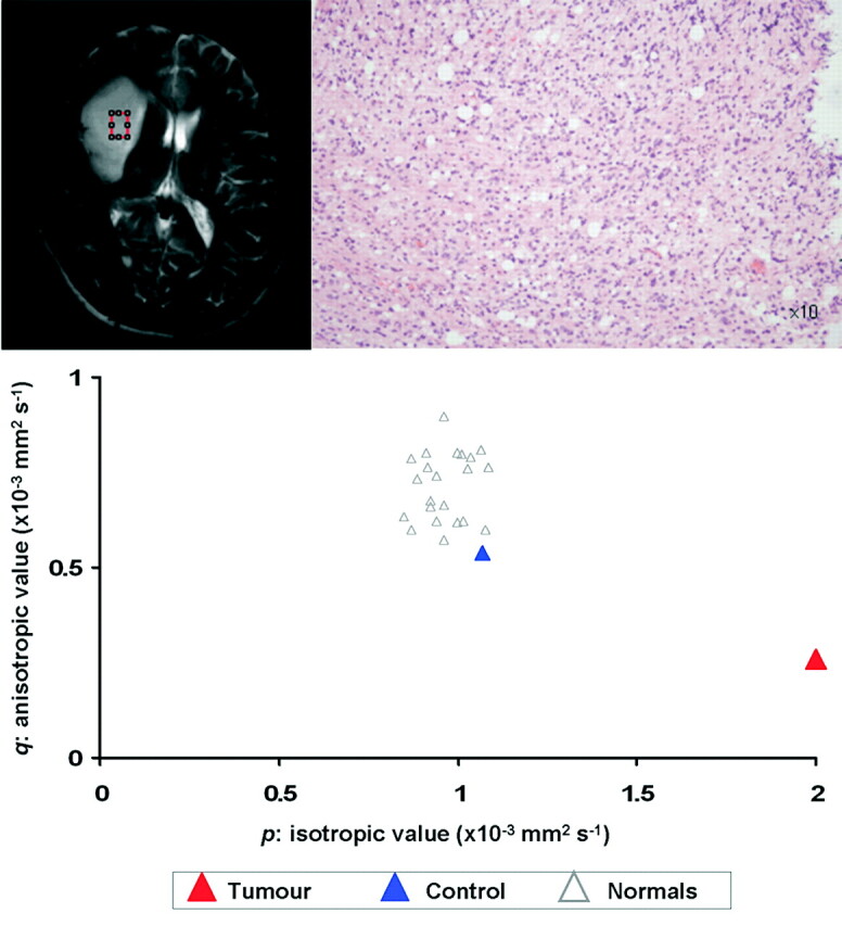
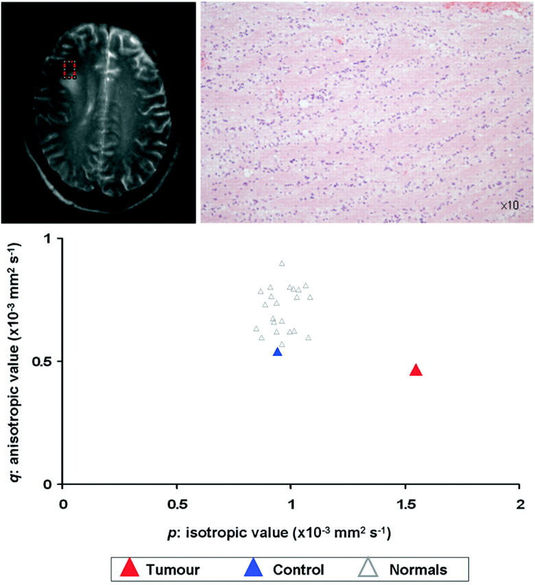
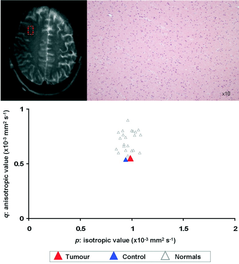
Results: Using diffusion tissue signatures, it was possible to differentiate gross tumor (reduction of the anisotropic component, q > 12% from contralateral region), from tumor infiltration (increase in the isotropic component, p > 10% from contralateral region). This technique has a sensitivity of 98% and specificity of 81%. T2-weighted abnormalities failed to identify the margin in half of all specimens.
Conclusion: Diffusion tensor imaging can better delineate the tumor margin in gliomas. Such techniques can improve the delineation of the radiation therapy target volume for gliomas and potentially can direct local therapies for tumor infiltration.
Figures
References
-
- Walker MD, Alexander E Jr, Hunt WE, et al. Evaluation of BCNU and/or radiotherapy in the treatment of anaplastic gliomas. A cooperative clinical trial. J Neurosurg 1978;49:333–43 - PubMed
-
- Oppitz U, Maessen D, Zunterer H, et al. 3D-recurrence-patterns of glioblastomas after CT-planned postoperative irradiation. Radiother Oncol 1999;53:53–57 - PubMed
-
- Chan JL, Lee SW, Fraass BA, et al. Survival and failure patterns of high-grade gliomas after three-dimensional conformal radiotherapy. J Clin Oncol 2002;20:1635–42 - PubMed
-
- Fitzek MM, Thornton AF, Rabinov JD, et al. Accelerated fractionated proton/photon irradiation to 90 cobalt gray equivalent for glioblastoma multiforme: results of a phase II prospective trial. J Neurosurg 1999;91:251–60 - PubMed
-
- Daumas-Duport C, Meder JF, Monsaingeon V, et al. Cerebral gliomas: malignancy, limits and spatial configuration. Comparative data from serial stereotaxic biopsies and computed tomography (a preliminary study based on 50 cases). J Neuroradiol 1983;10:51–80 - PubMed
MeSH terms
Grants and funding
LinkOut - more resources
Full Text Sources
Other Literature Sources
Medical
Research Materials