

Diffusion anisotropy changes in the brains of professional boxers
- PMID: 17032883
- PMCID: PMC7977918
Diffusion anisotropy changes in the brains of professional boxers
Abstract
Background and purpose: Professional boxing may result in brain injury. We hypothesize that quantitative MR diffusion imaging may be useful in determining early white matter changes.
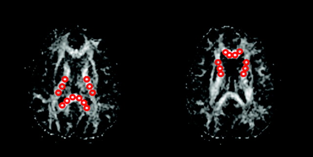
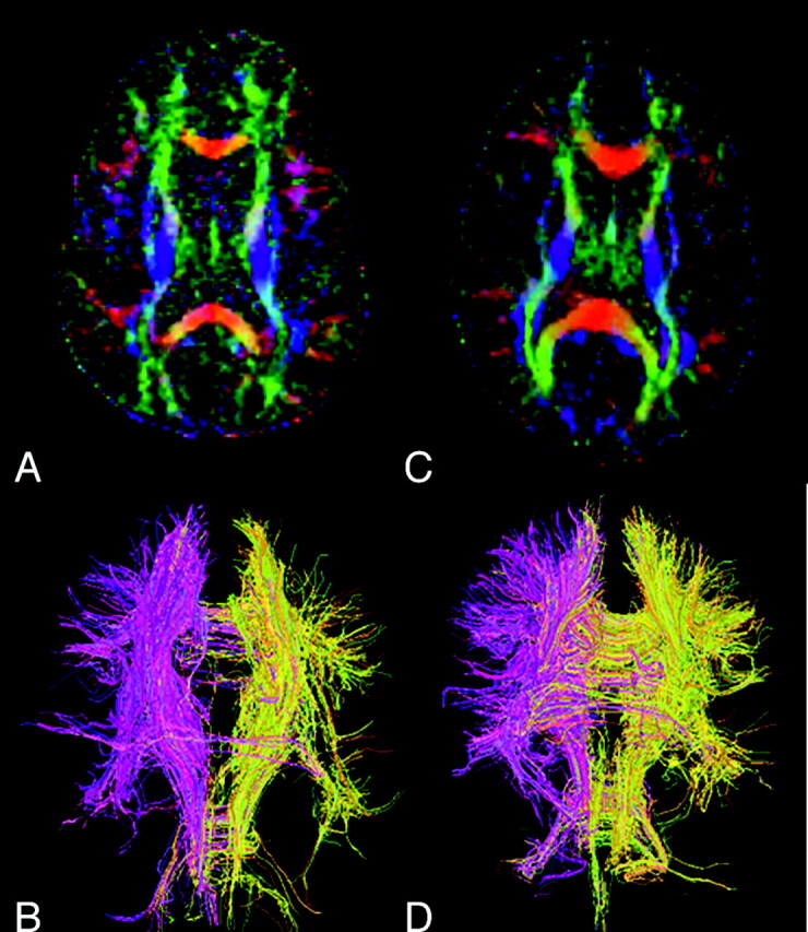
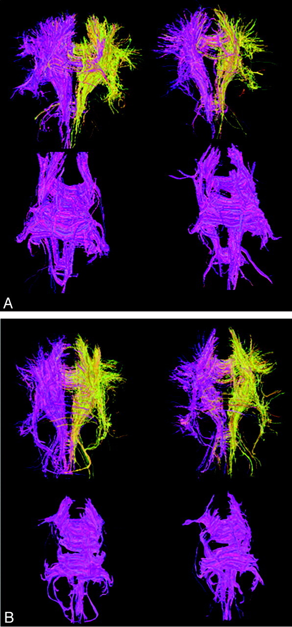
Methods: Forty-nine professional boxers (age 30 +/- 4.5 years) and 19 healthy control subjects (age 32 +/- 9.5 years) were imaged on a clinical 1.5T scanner. None of the subjects had neurologic disorder or deficit. The average diffusion constant (D(av)) and diffusion anisotropy (FA) were determined pixel by pixel. Regional diffusion measurements were done in the corpus callosum (CC) and internal capsule (IC). The whole brain diffusion constant (BD(av)) was also determined. Student t test was used to analyze the diffusion difference between boxers and the healthy control subjects. P < .05 was considered statistically significant.
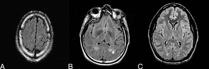
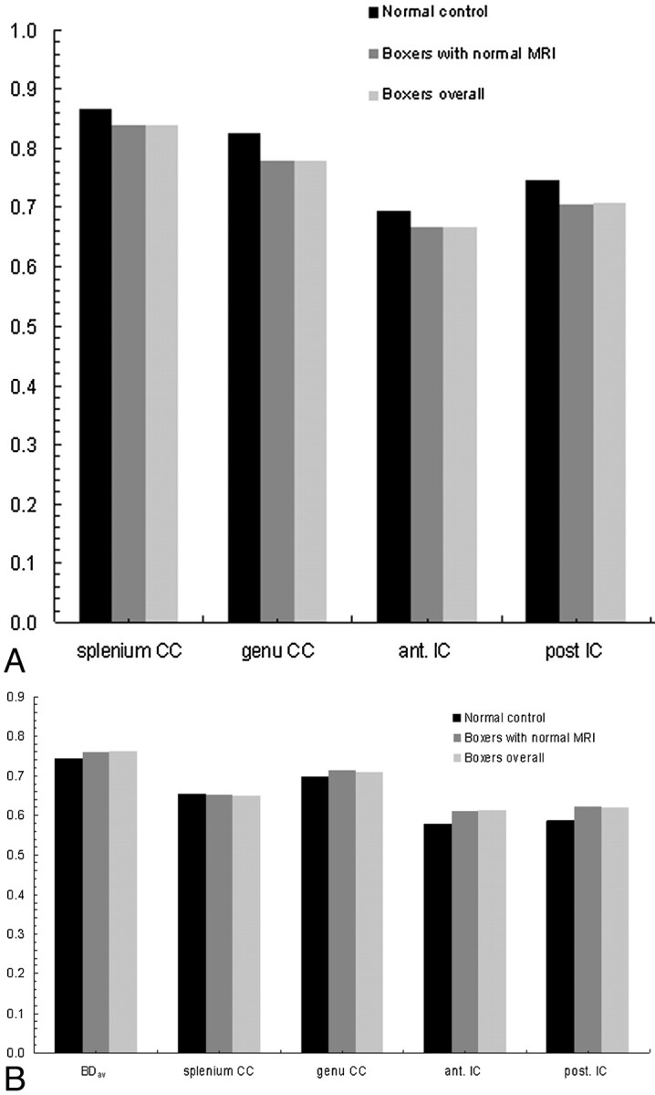
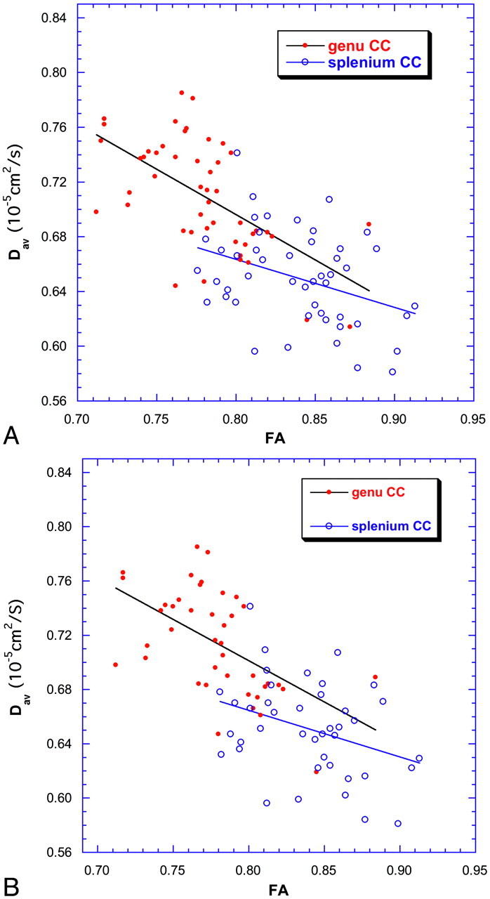
Results: Of the 49 professional boxers, 42 had normal conventional MRIs. The remaining 7 boxers had abnormal MR imaging findings dominated by nonspecific white matter disease. There was a significant difference in diffusion and anisotropy measurements in all the boxers compared with the healthy control subjects. In the boxer group, BD(av) increased and FA decreased significantly in the CC and posterior limb of IC. The measured FA and D(av) inversely correlated in regions of CC and IC in boxers but not in healthy control subjects. BD(av) also robustly correlated with both FA and D(av) in the splenium of CC in boxers.
Conclusion: Increased BD(av) and the decreased FA in the CC and IC may represent preclinical signs of subtle brain injury in professional boxers.
Figures
References
-
- Ross RJ, Casson JR, Siegel O, et al. Boxing injuries: neurologic, radiologic and neuropsychologic evaluation. Clin Sports Med 1987;6:41–51 - PubMed
-
- Jordan BD, Relkin NR, Ravdin LD, et al. Apolipoprotein E epsilon4 associated with chronic traumatic brain injury in boxing. JAMA 1997;278:136–40 - PubMed
-
- Moseley IF. The neuroimaging evidence for chronic brain damage due to boxing. Neuroradiology 2000;42:1–8 - PubMed
-
- Rodriguez G, Vitali P, Nobili F. Long-term effects of boxing and judochoking techniques on brain function. Ital J Neurol Sci 1998;19:367–72 - PubMed
MeSH terms
LinkOut - more resources
Full Text Sources
Other Literature Sources
Medical