. 2007 Feb 15;109(4):1736-42.
doi: 10.1182/blood-2006-03-010413. Epub 2006 Oct 10.

The impact of altered p53 dosage on hematopoietic stem cell dynamics during aging

Affiliations

The impact of altered p53 dosage on hematopoietic stem cell dynamics during aging

Melissa Dumble et al. Blood. .

Abstract

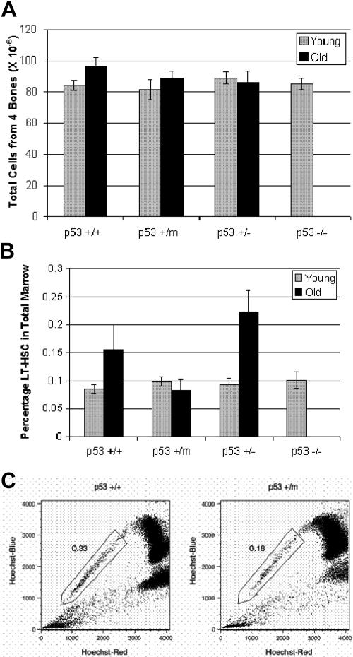
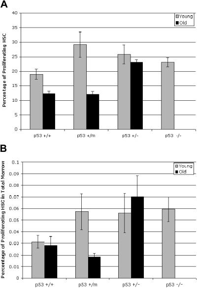
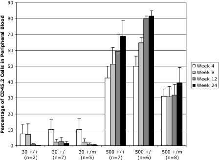
A temporal decline in tissue stem cell functionality may be a key component of mammalian aging. The tumor suppressor p53 has recently been implicated as a potential regulator of aging. We examined age-associated hematopoietic stem cell (HSC) dynamics in mice with varying p53 activities. Reduced p53 activity in p53+/- mice was associated with higher numbers of proliferating hematopoietic stem and progenitor cells in old age compared with aged wild-type (p53+/+) mice. We also assessed HSC dynamics in a p53 mutant mouse model (p53+/m) with higher apparent p53 activity than wild-type mice. The p53 hypermorphic (p53+/m) mice display phenotypes of premature aging. Many aged p53+/m organs exhibit reduced cellularity and atrophy, suggesting defects in stem-cell regenerative capacity. HSC numbers from old p53+/m mice fail to increase with age, unlike those of their p53+/+ and p53+/- counterparts. Moreover, transplantation of 500 HSCs from old p53+/m mice into lethally irradiated recipients resulted in reduced engraftment compared with old wild-type p53+/+ and p53+/- HSCs. Thus, alteration of p53 activity affects stem-cell numbers, proliferation potential, and hematopoiesis in older organisms, supporting a model in which aging is caused in part by a decline in tissue stem cell regenerative function.


Figures

Figure 1
Figure 1
Hematopoietic stem cells exhibit p53-dependent differences in stem-cell numbers in old mice. (A) Numbers of total bone marrow cells from young and old p53+/+, p53+/m, p53+/−, and p53−/− mice were obtained from hind limbs, counted manually and shown to be similar. Results represented are the mean ± SE of 10 to 14 mice per age/genotype. (B) LT-HSCs identified as lin−/lo, Sca-1+, c-kit+, and Flk-2 were selected by whole bone marrow antibody staining and subsequent flow cytometry. The experiments were performed in triplicate; n = 10 for young mice and n = 6 for old mice. Results shown represent the mean ± SE of the experiments. An increase in LT-HSC numbers is shown with age in p53+/+ and p53+/− bone marrow but not in p53+/m bone marrow. (C) Whole bone marrow was stained with Hoechst 33342 and enriched for Sca-1+ cells to identify the side population (SP) HSCs. SP-HSCs isolated from 12-month p53+/m bone marrow are reduced approximately 50% in number compared with SP-HSCs isolated from 12-month p53+/+ bone marrow. Numbers shown are for Sca-1+–enriched bone marrow, which enriches approximately 10-fold for SP cells.
Figure 2
Figure 2
Hematopoietic stem cells display p53-dependent differences in proliferation in old mice. (A) Whole bone marrow was labeled with BrdU for 16 hours before numbers of BrdU+, lin−/lo, Sca-1+, or c-kit+ HSCs were assessed by flow cytometry. BrdU incorporation was reduced in HSCs obtained from old p53+/+ and p53+/m mice but not in HSCs from old p53+/− mice. Results shown represent the mean ± SE of 2 experiments using a total of 5 (young) or 7 (old) mice. (B) The fraction of proliferating HSCs in the bone marrow of old mice is dependent on p53 status. The number of HSCs for each p53 genotype in Figure 2B was combined with the fraction of proliferating HSCs for each p53 genotype to determine the total number of proliferating HSCs in the bone marrow for each p53 genotype.
Figure 3
Figure 3
Limiting-dilution transplantation of 500 LT-HSCs (but not 30 HSCs) from old wild-type and p53 mutant mice results in differential white blood cell reconstitution in lethally irradiated recipients. LT-HSCs were isolated from CD45.2 donor mice based on their status as lin−/lo, Sca-1+, c-kit+, Flk-2. Either 30 or 500 LT-HSCs from CD45.2 test mice were injected into marrow-ablated CD45.1 recipients. Recipient mice were analyzed 4, 8, 12, and 24 weeks after transplantation to assess the percentage of peripheral white blood cells produced from transplanted CD45.2 LT-HSCs. These experiments were performed in duplicate. For each experiment, 3 old donor mice of the same genotype were used, donor cells were pooled, and HSCs were isolated and injected into 6 irradiated recipient mice. Error bars indicate standard error of the mean.

References

    1. Weissman IL. Stem cells: units of development, units of regeneration, and units in evolution. Cell. 2000;100:157–168. - PubMed
    1. Van Zant G, Liang Y. The role of stem cells in aging. Exp Hematol. 2003;31:659–672. - PubMed
    1. Chen J. Senescence and functional failure in hematopoietic stem cells. Exp Hematol. 2004;32:1025–1032. - PubMed
    1. Sharpless NE, DePinho RA. Telomeres, stem cells, senescence, and cancer. J Clin Invest. 2004;113:160–168. - PMC - PubMed
    1. Pelicci PG. Do tumor-suppressive mechanisms contribute to organism aging by inducing stem cell senescence? J Clin Invest. 2004;113:4–7. - PMC - PubMed

Publication types

Substances