Case Reports
. 2006 Nov;38(11):1310-5.
doi: 10.1038/ng1905. Epub 2006 Oct 8.

Loss of DMP1 causes rickets and osteomalacia and identifies a role for osteocytes in mineral metabolism

Affiliations
Case Reports

Loss of DMP1 causes rickets and osteomalacia and identifies a role for osteocytes in mineral metabolism

Jian Q Feng et al. Nat Genet. 2006 Nov.

Abstract

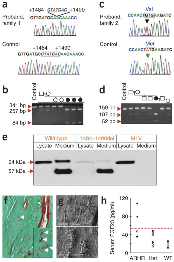
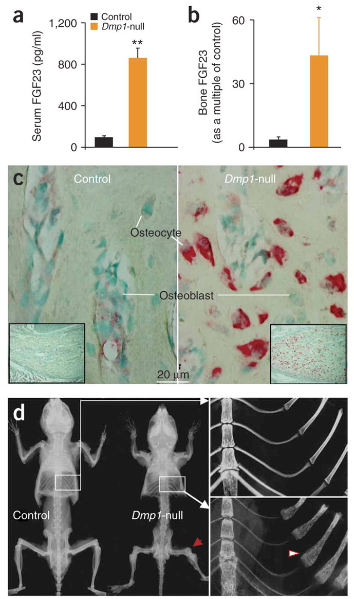
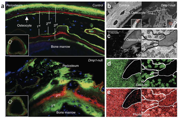
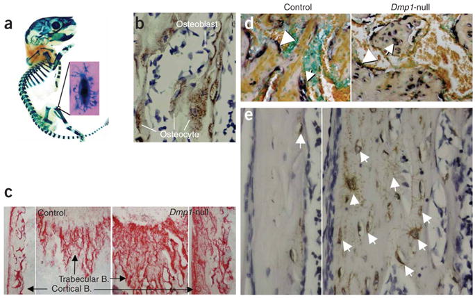
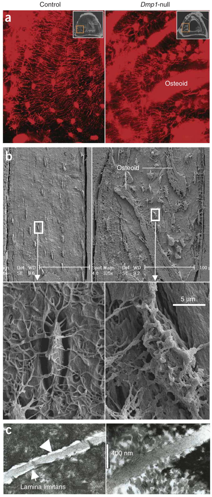
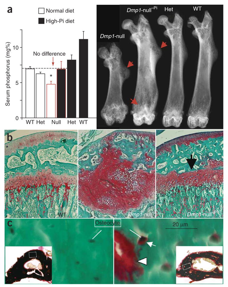
The osteocyte, a terminally differentiated cell comprising 90%-95% of all bone cells, may have multiple functions, including acting as a mechanosensor in bone (re)modeling. Dentin matrix protein 1 (encoded by DMP1) is highly expressed in osteocytes and, when deleted in mice, results in a hypomineralized bone phenotype. We investigated the potential for this gene not only to direct skeletal mineralization but also to regulate phosphate (P(i)) homeostasis. Both Dmp1-null mice and individuals with a newly identified disorder, autosomal recessive hypophosphatemic rickets, manifest rickets and osteomalacia with isolated renal phosphate-wasting associated with elevated fibroblast growth factor 23 (FGF23) levels and normocalciuria. Mutational analyses showed that autosomal recessive hypophosphatemic rickets family carried a mutation affecting the DMP1 start codon, and a second family carried a 7-bp deletion disrupting the highly conserved DMP1 C terminus. Mechanistic studies using Dmp1-null mice demonstrated that absence of DMP1 results in defective osteocyte maturation and increased FGF23 expression, leading to pathological changes in bone mineralization. Our findings suggest a bone-renal axis that is central to guiding proper mineral metabolism.


Figures

Figure 1
Figure 1
DMP1 mutations, osteomalacia and a defective osteocyte lacunocanalicular network in ARHR. (a) Family 1 had a biallelic deletion of nucleotides 1484–1490 in DMP1 exon 6 (missing nucleotides outlined). (b) The 1484–1490del segregates with the disorder, as assessed by RFLP, creating a new HpyCH4V site, which creates 257- and 84-bp fragments from the 341-bp exon 6 PCR product (circles: female; square: male, filled symbol: affected individuals; individuals F1-1, F1-2 and F1-3 are depicted chronologically from left to right). (c) Family 2 had a start codon mutation (A1→G) resulting in a methionine to valine change (M1V) not present in control individuals. (d) M1V segregates with the ARHR phenotype in family 2 and creates new 52-bp and 107-bp TaaI fragments from a 159-bp PCR product. (e) Wild-type (WT) and ARHR mutant DMP1 expression in HEK293 cells. Wild-type DMP1 was detected by protein blot analyses as a 94-kDa protein in the cellular lysates and as 94-kDa and 57-kDa polypeptides in the growth medium. The 1484–1490del mutant was primarily secreted as the 57-kDa form of DMP1, with fainter expression in the cellular lysates, whereas the M1V mutant was retained within the cell as the 94-kDa form of DMP1 and had no detectable signal in the medium. (f) Goldner staining indicates abundant osteoid (red) on bone edges (arrowheads) and surrounding osteocytes (arrows). (g) Resin-casted SEM images show osteocyte lacunae in a cluster (top), with few dendrites and rough surfaces (bottom). (h) Serum FGF23 levels: ARHR individuals compared with heterozygous (Het) and wild-type individuals. Filled circles: family 1; open circles: family 2. Dotted line represents upper limit of normal (54 pg/ml).
Figure 2
Figure 2
Dmp1-null mice show skeletal abnormalities, rickets and elevated FGF23. (a) Serum Fgf23 levels are shown for Dmp1-null mice compared with control heterozygote littermates. Data are mean ± s.e.m. from 2- to 5-month-old mice; n = 6 (Dmp1-null), n = 11 (control); **P < 0.01. (b) Real time RT-PCR of Dmp1-null long bone demonstrates marked elevation of Fgf23 mRNA expression (*P < 0.05). (c) In situ hybridization shows increased Fgf23 mRNA expression (red) in 10-d-old Dmp1-null osteocytes only. (d) Representative radiographs of skeletons from control and Dmp1-null mice at 3 months of age. In the Dmp1-null skeleton, the flared ends of long bones are indicated by arrows and the rachitic rosary of the ribs by an arrowhead.
Figure 3
Figure 3
Dmp1-null mice show defects in mineralization. (a) Confocal microscopy images of fluorochrome labeling, counterstained with DAPI for visualization of osteocyte nuclei (blue). Dmp1- null osteocytes are buried in diffuse fluorochrome label, suggesting a defect in the process of mineral propagation. (b) Images of backscattered EM of tibias from 6-week-old mice (samples were treated with osmium to preserve cell morphology). (ce) STEM maps of unstained osmium-free thin sections (<1 mm) from the same tibias of control (left) and Dmp1-null mice (right). With this technology, the convergent electron beam is scanned over a defined area of the sample to obtain mineral (c, black), calcium (d, green) and phosphorus (e, red/white) distribution within matrix.
Figure 4
Figure 4
Defective osteoblast-to-osteocyte differentiation and maturation in Dmp1-null mice. (a) A whole mount X-gal stain of a skeleton from an 8-day-old Dmp1-lacZ knock-in pup. (b) DMP1 immunostain of bone matrix surrounding osteocytes. (c) An increase in alkaline phosphatase activity in 10-d-old Dmp1-null bone matrix (right). Arrows indicate region of magnification. B = bone. (d) Abnormal expression of type 1 collagen mRNA in Dmp1-null osteocytes. Arrows indicate osteocytes; arrowheads indicate osteoblasts. (e) Highly expressed E11 protein in all Dmp1-null osteocytes.
Figure 5
Figure 5
Defective organization of osteocyte lacunae and lacunocanalicular walls in Dmp1-null mice. (a) Visualization of disorganized osteocyte-canalicular system in Dmp1-null mice with procion red injection compared with the well-organized control osteocytes (left) using confocal microscopy at 40× at 565 nm excitation and 610 nm emission. (b) SEM images of the acid-etched, resin-casted osteocyte-canalicular system. Note the differences between the control (left) and the Dmp1-null (right) in distribution, size and surface of osteocytes. (c) TEM images of sagittal sections of osteocyte canaliculi and dendrites (control, left; Dmp1-null, right).
Figure 6
Figure 6
High-phosphate diet rescues the rickets but not the osteomalacic feature of the Dmp1-null phenotype. (a) Restoration of phosphate homeostasis by high-phosphate diet for 4 weeks (left) leads to rescue of rickets in Dmp1-null mice as shown by autoradiography (right). mg% = 1 mg/100 ml. Dmp1-null−Pi indicates an animal on a high-phosphate diet. ‘Het’ = heterozygote. (b) Confirmation of rickets rescue using safranin-O staining of growth plates. (c) High-phosphate diet has a limited effect on the Dmp1-null osteomalacia phenotype. Goldner stain shows abundant osteoid (red) is still present on bone edges (arrowhead) and surrounding osteocytes (arrow). Goldner staining is validated by von Kossa staining (black, mineral; red, osteoid; insets show low magnification). *Indicates statistically significant difference (P < 0.05).

Comment in

  • Bone talk.
    Schiavi SC. Schiavi SC. Nat Genet. 2006 Nov;38(11):1230-1. doi: 10.1038/ng1106-1230. Nat Genet. 2006. PMID: 17072297 No abstract available.

References

    1. Frost HM. In vivo osteocyte death. J Bone Joint Surg Am. 1960;42A:138–143. - PubMed
    1. Palumbo C, Palazzini S, Zaffe D, Marotti G. Osteocyte differentiation in the tibia of newborn rabbit: an ultrastructural study of the formation of cytoplasmic processes. Acta Anat (Basel) 1990;137:350–358. - PubMed
    1. Pead MJ, Lanyon LE. Indomethacin modulation of load-related stimulation of new bone formation in vivo. Calcif Tissue Int. 1989;45:34–40. - PubMed
    1. Toyosawa S, et al. Dentin matrix protein 1 is predominantly expressed in chicken and rat osteocytes but not in osteoblasts. J Bone Miner Res. 2001;16:2017–2026. - PubMed
    1. Ling Y, et al. DMP1 depletion decreases bone mineralization in vivo: an FTIR imaging analysis. J Bone Miner Res. 2005;20:2169–2177. - PMC - PubMed

Publication types

MeSH terms