. 2006 Nov;79(5):869-77.
doi: 10.1086/508434. Epub 2006 Sep 15.

Distinct clinical phenotypes associated with a mutation in the mitochondrial translation elongation factor EFTs

Affiliations

Distinct clinical phenotypes associated with a mutation in the mitochondrial translation elongation factor EFTs

Jan A M Smeitink et al. Am J Hum Genet. 2006 Nov.

Abstract

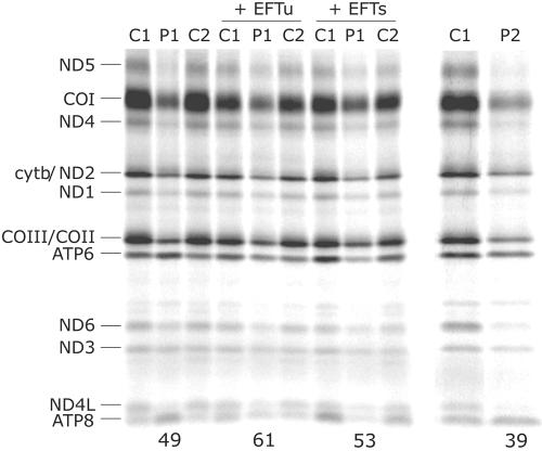
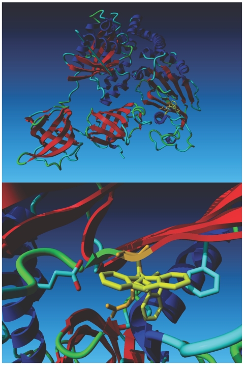
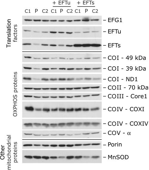
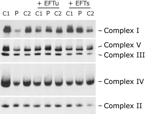
The 13 polypeptides encoded in mitochondrial DNA (mtDNA) are synthesized in the mitochondrial matrix on a dedicated protein-translation apparatus that resembles that found in prokaryotes. Here, we have investigated the genetic basis for a mitochondrial protein-synthesis defect associated with a combined oxidative phosphorylation enzyme deficiency in two patients, one of whom presented with encephalomyopathy and the other with hypertrophic cardiomyopathy. Sequencing of candidate genes revealed the same homozygous mutation (C997T) in both patients in TSFM, a gene coding for the mitochondrial translation elongation factor EFTs. EFTs functions as a guanine nucleotide exchange factor for EFTu, another translation elongation factor that brings aminoacylated transfer RNAs to the ribosomal A site as a ternary complex with guanosine triphosphate. The mutation predicts an Arg333Trp substitution at an evolutionarily conserved site in a subdomain of EFTs that interacts with EFTu. Molecular modeling showed that the substitution disrupts local subdomain structure and the dimerization interface. The steady-state levels of EFTs and EFTu in patient fibroblasts were reduced by 75% and 60%, respectively, and the amounts of assembled complexes I, IV, and V were reduced by 35%-91% compared with the amounts in controls. These phenotypes and the translation defect were rescued by retroviral expression of either EFTs or EFTu. These data clearly establish mutant EFTs as the cause of disease in these patients. The fact that the same mutation is associated with distinct clinical phenotypes suggests the presence of genetic modifiers of the mitochondrial translation apparatus.


Figures

Figure  1.
Figure 1.
Pulse labeling of mitochondrial translation products in patient fibroblasts. Fibroblasts from the two patients (P1 and P2) and two controls (C1 and C2) were labeled with [35S]methionine in the presence of emetine, a cytosolic translation inhibitor, as outlined in the “Material and Methods” section. Fibroblasts from patient 1 were also analyzed after transduction with retroviral vectors overexpressing the mitochondrial translation factors EFTu or EFTs. The mitochondrial translation products are indicated on the left of the autoradiogram. COI–COIII = subunits of cytochrome c oxidase; cytb = cytochrome b subunit; ND1, ND2, ND3, ND4, ND4L, ND5, and ND6 = subunits of NADH CoQ oxidoreductase. The numbers at the bottom of the gel indicate the total incorporation of [35S]methionine relative to the corresponding control.
Figure  2.
Figure 2.
Sequence analysis of TSFM. A, DNA sequence chromatogram from a control and patient 1 (homozygous C997T) and a heterozygous chorionic villus sample. B, Domain structure of EFTs (N-terminal in black, subdomain N in white, and subdomain C in gray) showing the predicted Arg333Trp substitution in subdomain. C, Protein sequence alignment for Homo sapiens, Bos taurus, Caenorhabditis elegans, and E. coli. The alignment from human to bacteria shows that the mutated Arg333Trp is highly conserved among different species. D, Analysis of microsatellite markers spanning the TSFM locus on chromosome 12. The markers are arbitrarily assigned to chromosomes in both patients.
Figure  3.
Figure 3.
Computational modeling based on the crystal structure of the bovine EFTu•EFTs complex. The top panel shows the structure of the Tu•Ts dimer. Tu is on the left side of the model, and Ts is on the right. The wild-type Arg 333 residue (depicted in orange) is superimposed on three Trp rotamers (depicted in yellow). The lower panel is a close-up of the region containing the mutation, which shows that the Arg residue fits well in front of the C-terminal side of the helix beneath it. The most favorable Trp rotamer bumps into this same helix. The second- and third-best local minima for the Trp rotamer bump into Glu 644 and Phe 637, respectively. The second rotamer also bumps into its own backbone.
Figure  4.
Figure 4.
Immunoblot analysis of fibroblasts from patient 1. Immunoblot analysis of the steady-state levels of three translation elongation factors (EFG1, EFTu, and EFTs) and eight OXPHOS system subunits (COI–49 kDa, COI–39 kDa, COI-ND1, COII–70 kDa, COIII-Core1, COIV-COXI, COIV-COXIV, and COV-α) in control and patient fibroblasts before and after transduction with retroviral constructs expressing the mitochondrial translation elongation factors EFTu or EFTs. Porin and manganese superoxide dismutase (MnSOD) were used as loading controls.
Figure  5.
Figure 5.
Blue-native PAGE analysis of the assembly of the OXPHOS complexes in patient fibroblasts. Fibroblasts from patient 1 and two controls (C1 and C2) were analyzed by blue-native PAGE before and after transduction with retroviral constructs expressing the mitochondrial translation elongation factors EFTu or EFTs. The gels were immunoblotted with antibodies directed against specific individual subunits to assess the amount of each of the fully assembled complexes.

References

Web Resources

    1. BLAST, http://www.ncbi.nlm.nih.gov/BLAST/
    1. GenBank, http://www.ncbi.nlm.nih.gov/Genbank/ (for TSFM [accession number NM_005726] and TUFM [accession number NM_003321])
    1. Helper Dependent Protocol, http://www.stanford.edu/group/nolan/protocols/pro_helper_dep.html
    1. Online Mendelian Inheritance in Man (OMIM), http://www.ncbi.nlm.nih.gov/Omim/ (for TSFM) - PubMed
    1. POV-Ray, http://www.povray.org/

References

    1. Thorburn DR (2004) Mitochondrial disorders: prevalence, myths and advances. J Inherit Metab Dis 27:349–362 10.1023/B:BOLI.0000031098.41409.55 - DOI - PubMed
    1. Smeitink J, van den Heuvel L, DiMauro S (2001) The genetics and pathology of oxidative phosphorylation. Nat Rev Genet 2:342–352 10.1038/35072063 - DOI - PubMed
    1. Jacobs HT (2003) Disorders of mitochondrial protein synthesis. Hum Mol Genet Spec No 2 12:R293–R301 10.1093/hmg/ddg285 - DOI - PubMed
    1. Spremulli LL, Coursey A, Navratil T, Hunter SE (2004) Initiation and elongation factors in mammalian mitochondrial protein biosynthesis. Prog Nucleic Acid Res Mol Biol 77:211–261 - PubMed
    1. Zhang Y, Spremulli LL (1998) Identification and cloning of human mitochondrial translational release factor 1 and the ribosome recycling factor. Biochim Biophys Acta 1443:245–250 - PubMed

Publication types

MeSH terms

Associated data

LinkOut - more resources