Case Reports
. 2006 Nov;79(5):878-89.
doi: 10.1086/508474. Epub 2006 Sep 6.

Mutations of the mitochondrial holocytochrome c-type synthase in X-linked dominant microphthalmia with linear skin defects syndrome

Affiliations
Case Reports

Mutations of the mitochondrial holocytochrome c-type synthase in X-linked dominant microphthalmia with linear skin defects syndrome

Isabella Wimplinger et al. Am J Hum Genet. 2006 Nov.

Abstract

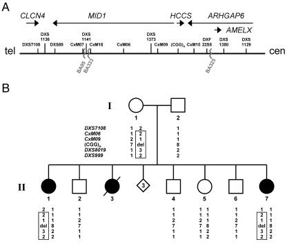
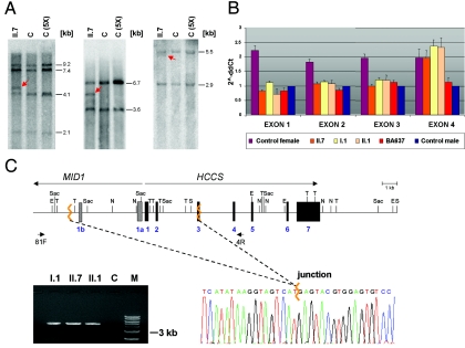
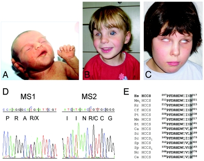
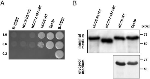
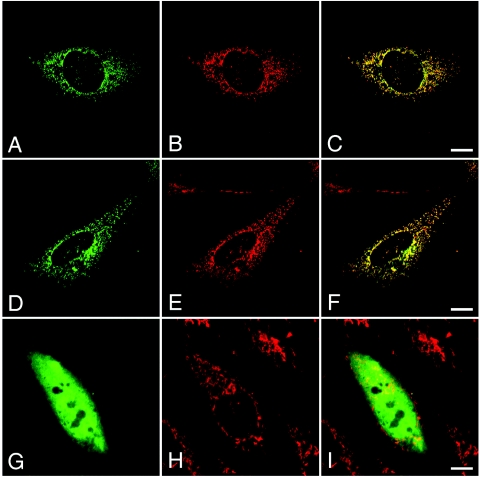
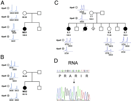
The microphthalmia with linear skin defects syndrome (MLS, or MIDAS) is an X-linked dominant male-lethal disorder almost invariably associated with segmental monosomy of the Xp22 region. In two female patients, from two families, with MLS and a normal karyotype, we identified heterozygous de novo point mutations--a missense mutation (p.R217C) and a nonsense mutation (p.R197X)--in the HCCS gene. HCCS encodes the mitochondrial holocytochrome c-type synthase that functions as heme lyase by covalently adding the prosthetic heme group to both apocytochrome c and c(1). We investigated a third family, displaying phenotypic variability, in which the mother and two of her daughters carry an 8.6-kb submicroscopic deletion encompassing part of the HCCS gene. Functional analysis demonstrates that both mutant proteins (R217C and Delta 197-268) were unable to complement a Saccharomyces cerevisiae mutant deficient for the HCCS orthologue Cyc3p, in contrast to wild-type HCCS. Moreover, ectopically expressed HCCS wild-type and the R217C mutant protein are targeted to mitochondria in CHO-K1 cells, whereas the C-terminal-truncated Delta 197-268 mutant failed to be sorted to mitochondria. Cytochrome c, the final product of holocytochrome c-type synthase activity, is implicated in both oxidative phosphorylation (OXPHOS) and apoptosis. We hypothesize that the inability of HCCS-deficient cells to undergo cytochrome c-mediated apoptosis may push cell death toward necrosis that gives rise to severe deterioration of the affected tissues. In summary, we suggest that disturbance of both OXPHOS and the balance between apoptosis and necrosis, as well as the X-inactivation pattern, may contribute to the variable phenotype observed in patients with MLS.


Figures

Figure  1.
Figure 1.
Minimal MLS critical region and haplotype analysis of the family of patient II.7. A, Schematic representation of the minimal MLS critical region (610 kb) in Xp22.2, defined by the breakpoints of patients BA333 and BA325 (jagged lines). The figure is not drawn to scale. The uppermost arrows show the orientation of genes within the critical region. The position of DNA marker loci previously mapped in this region is shown. B, Haplotype analysis of six representative polymorphic markers in Xp22.2-p22.13 in the family of patient II.7. Whereas markers DXS7108, CxM06, CxM09, and SNP rs5901444—(CGG)n—are positioned in the MLS critical region and indicated in panel A, loci DXS8019 and DXS999 are located centromeric to the critical region. The polymorphic (CGG)n repeat is located in the HCCS 5′ UTR, and the respective repeat length is given. Alleles are shown below the pedigree symbols. del = Deletion of one allele. The haplotype shared by the affected sisters (II.1 and II.7) and their mother (I.1) is boxed. Both patients II.7 and II.1 carry only the paternal allele, with eight CGG repeats, whereas the unaffected sister (II.5) is heterozygous for the trinucleotide repeat. Segregation analysis showed that the three brothers (II.2, II.4, and II.6) and the unaffected sister (II.5) carry a different maternal haplotype than do II.1 and II.7.
Figure  2.
Figure 2.
A submicroscopic deletion encompassing part of the HCCS gene is present in three members of the family of patient II.7. A, Southern blot hybridized with a probe covering the HCCS 5′ UTR and coding region. Genomic DNA samples of patient II.7 and controls were digested with SpeI and EcoRV (left), NdeI and SacI (middle), and TaqI (right). C = female control; C (5X) = human cell line with five X chromosomes. Various fragments of altered sizes (red arrows) were observed in II.7. B, Relative quantification of copy numbers of HCCS exons 1–4, by real-time PCR on genomic DNA of individuals I.1, II.1, and II.7. BA637 has MLS and displays monosomy of the Xp22 region. Values of HCCS exons 1–3 in II.7, II.1, and I.1 are comparable to that of a haploid sample. C, Schematic representation of part of the MLS critical region (top). MID1 and HCCS genes are indicated by long arrows, and primers used for junction fragment amplification are indicated by short arrows. Exons 1a and 1b of MID1 are represented by bars, exons of HCCS by black boxes. Jagged orange lines indicate deletion breakpoints. E = EcoRV; N = NdeI; S = SpeI; Sac = SacI; T = TaqI. Bottom left panel, Amplification of a junction fragment from genomic DNA of I.1, II.1, and II.7 but not from a control (C). M = DNA marker. Bottom right panel, Part of the DNA sequence electropherogram of the rearrangement-specific junction fragment. The deletion breakpoint is indicated by a jagged orange line. The 8.6-kb deletion encompasses HCCS exons 1–2, part of exon 3, and exons 1a and 1b of MID1.
Figure  3.
Figure 3.
Point mutations in HCCS in two patients with MLS. A–C, Photographs of patients MS1 and MS2. A, Linear skin defects located on the face and neck of patient MS1 at birth. B, Microphthalmia of the right eye and bilateral sclerocornea of patient MS1 at age 4 years. Linear skin defects have completely disappeared. C, Patient MS2, at age 8 years, with bilateral microphthalmia and sclerocornea. No linear skin defects were noted at birth. D, Sequence electropherograms of part of HCCS exons from genomic DNA of patients MS1 and MS2. Nucleotide triplets and encoded amino acids are indicated. Patient MS1 is heterozygous for the c.589C→T mutation (p.R197X) (left panel) in exon 6, whereas the heterozygous mutation c.649C→T (p.R217C) (right panel) in exon 7 was found in patient MS2. E, Partial amino acid–sequence alignment of heme lyases from various species. The position of amino acids is given. Evolutionarily conserved residues are shown in bold. The invariant arginine at position 217, which is altered to cysteine in patient MS2, is shaded in gray. HCCS1 indicates specificity of the heme lyase for cytochrome c1. Hs = Homo sapiens; Mm1 = Mus musculus; Rr = Rattus norvegicus; Cf = Canis familiaris; Pt = Pan troglodytes; Mm = Macaca mulatta; Bt = Bos taurus; Ca = Candida albicans; Sc = S. cerevisiae; Sp = Schizosaccharomyces pombe; Nc = Neurospora crassa; Ce = Caenorhabditis elegans.
Figure  4.
Figure 4.
HCCS mutant proteins are not able to complement S. cerevisiae CYC3 deficiency. A, Functional complementation of the S. cerevisiae strain B-8025. B-8025 was transformed with human wild-type HCCS (HCCS WT), the mutants Δ197–268 and R217C, or yeast CYC3 (Cyc3p) expression constructs and was grown on minimal medium. Transformants were grown in liquid minimal medium, and aliquots of 5 μl of saturated and diluted cultures were spotted on glycerol medium containing copper (to induce expression of GST-fusion proteins) and were incubated for 5 d at 30°C. The top row shows spots of saturated cultures, and the middle and bottom rows show spots of dilutions; dilution rates are indicated to the left of the figure. Note partial restoration of growth by Cyc3p and wild-type HCCS, whereas no growth was observed for the untransformed strain or that expressing HCCS Δ197–268 or HCCS R217C. In parallel, all strains were also spotted on plates with glucose-containing minimal medium and showed normal growth (data not shown). Strain B-7553 served as wild-type growth control. B, Expression of GST-HCCS–fusion proteins determined by western blotting. Expression of GST-HCCS R217C–, Δ197–268–, wild-type–, and GST-Cyc3p–fusion proteins in yeast strain B-8025, grown in minimal medium with copper, was demonstrated by immunoblotting (top panel), whereas, in strain B-8025 grown in glycerol-containing medium, only GST-HCCS wild-type– and GST-Cyc3p–fusion proteins were expressed (lower panel). B-8025 transformed with the GST-HCCS Δ197–268 or GST-HCCS R217C construct did not grow under this condition.
Figure  5.
Figure 5.
Targeting of ectopically expressed HCCS wild-type and mutant proteins to mitochondria. Subcellular localization of different N-terminally HA-tagged HCCS proteins ectopically expressed in CHO-K1 cells (A, D, and G) and staining of endogenous mitochondria by MitoTracker (B, E, and H) are shown. HA-tagged HCCS wild-type protein (A [green]) is targeted to mitochondria (B [red]), as shown by colocalization with the MitoTracker (C [yellow]). Similarly, HA-tagged HCCS R217C mutant protein (D [green]) shows a mitochondrial (E [red]) distribution (F [yellow]). In contrast, the truncated HCCS Δ197–268 protein is diffusively dispersed in the cell (G), and the two fluorescence patterns (G and H) show no overlap (I). The scale bars represent 10 μm.
Figure  6.
Figure 6.
Skewed X inactivation in females with an HCCS mutation. X-chromosome inactivation determined by amplification of an AR-sequence polymorphism and digestion of genomic DNA isolated from lymphocytes with HpaII (indicated with plus [+] and minus [−] signs, respectively). The ratio of the X-inactivation pattern is given below the respective pedigree symbol. All females carrying an HCCS mutation show nonrandom or extremely skewed X inactivation (patient MS1 in panel A; patient MS2 in panel B; and II.1, II.7, and their mother [I.1] in panel C). D, Part of the DNA sequence electropherogram of HCCS exon 6, obtained from an RT-PCR amplicon of patient MS1. Only the wild-type allele (c.589C) (arrow) is expressed in her lymphoblastoid cells.

References

Web Resources

    1. GenBank, http://www.ncbi.nlm.nih.gov/Genbank/ (for HCCS [accession number NM_005333], RPP30 [accession number NM_006413], and HCCS cDNA clone DKFZp779I1858 [accession number CR749578])
    1. Online Mendelian Inheritance in Man (OMIM), http://www.ncbi.nlm.nih.gov/Omim/ (for MLS phenotypes)
    1. RZPD German Resource Center for Genome Research, http://www.rzpd.de/

References

    1. Van den Veyver IB (2002) Microphthalmia with linear skin defects (MLS), Aicardi, and Goltz syndromes: are they related X-linked dominant male-lethal disorders? Cytogenet Genome Res 99:289–296 10.1159/000071606 - DOI - PubMed
    1. Bird LM, Krous HF, Eichenfield LF, Swalwell CI, Jones MC (1994) Female infant with oncocytic cardiomyopathy and microphthalmia with linear skin defects (MLS): a clue to the pathogenesis of oncocytic cardiomyopathy? Am J Med Genet 53:141–148 10.1002/ajmg.1320530205 - DOI - PubMed
    1. Happle R, Daniels O, Koopman RJ (1993) MIDAS syndrome (microphthalmia, dermal aplasia, and sclerocornea): an X-linked phenotype distinct from Goltz syndrome. Am J Med Genet 47:710–713 10.1002/ajmg.1320470525 - DOI - PubMed
    1. Kherbaoui-Redouani L, Eschard C, Bednarek N, Morville P (2003) [Cutaneous aplasia, non compaction of the left ventricle and severe cardiac arrhythmia: a new case of MLS syndrome (microphthalmia with linear skin defects)]. Arch Pediatr 10:224–226 10.1016/S0929-693X(03)00014-9 - DOI - PubMed
    1. Morleo M, Pramparo T, Perone L, Gregato G, Le Caignec C, Mueller RF, Ogata T, Raas-Rothschild A, de Blois MC, Wilson LC, Zaidman G, Zuffardi O, Ballabio A, Franco B (2005) Microphthalmia with linear skin defects (MLS) syndrome: clinical, cytogenetic, and molecular characterization of 11 cases. Am J Med Genet A 137:190–198 - PubMed

Publication types

MeSH terms

Associated data