. 2006 Nov;79(5):949-57.
doi: 10.1086/508617. Epub 2006 Sep 19.

Mutations in the tight-junction gene claudin 19 (CLDN19) are associated with renal magnesium wasting, renal failure, and severe ocular involvement

Affiliations

Mutations in the tight-junction gene claudin 19 (CLDN19) are associated with renal magnesium wasting, renal failure, and severe ocular involvement

Martin Konrad et al. Am J Hum Genet. 2006 Nov.

Abstract

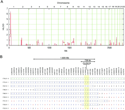
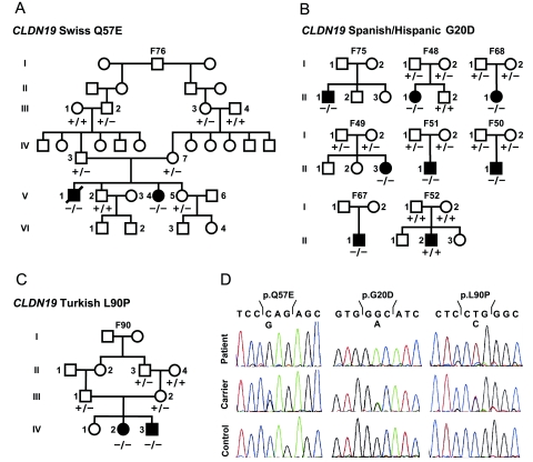
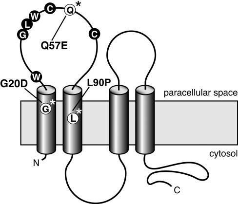
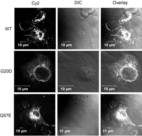
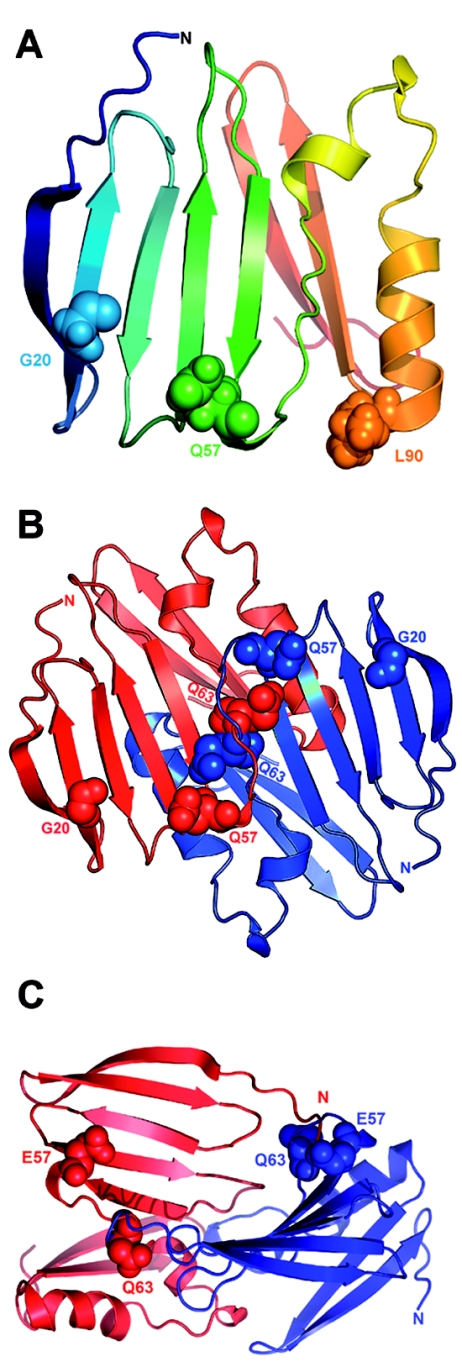
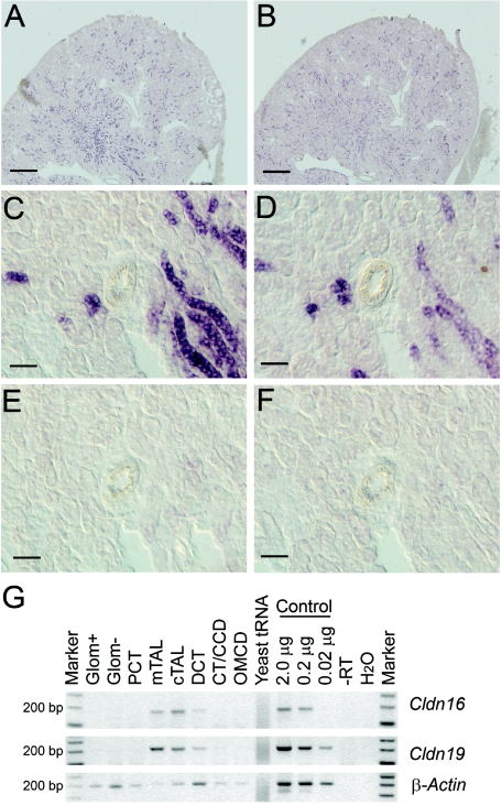
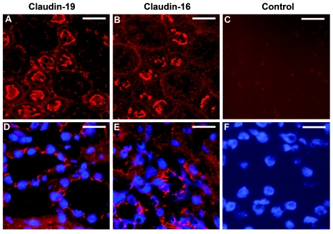
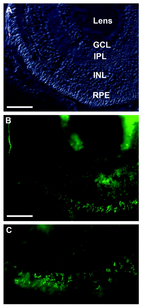
Claudins are major components of tight junctions and contribute to the epithelial-barrier function by restricting free diffusion of solutes through the paracellular pathway. We have mapped a new locus for recessive renal magnesium loss on chromosome 1p34.2 and have identified mutations in CLDN19, a member of the claudin multigene family, in patients affected by hypomagnesemia, renal failure, and severe ocular abnormalities. CLDN19 encodes the tight-junction protein claudin-19, and we demonstrate high expression of CLDN19 in renal tubules and the retina. The identified mutations interfere severely with either cell-membrane trafficking or the assembly of the claudin-19 protein. The identification of CLDN19 mutations in patients with chronic renal failure and severe visual impairment supports the fundamental role of claudin-19 for normal renal tubular function and undisturbed organization and development of the retina.


Figures

Figure  1.
Figure 1.
Mapping of a new gene locus for recessive renal hypomagnesemia on 1p34.2. A, Application of a reduced subset of 7,802 informative autosomal SNPs. The highest (by far) HLOD score was identified on chromosome 1p34.2, with a peak value of 6.4. Physical positions of the SNP markers are depicted on the abscissa. B, A critical interval of <2 Mb, revealed by comparison of the patients’ haplotypes. The position of the candidate gene CLDN19 and the flanking SNP markers (highlighted in yellow) are depicted. Homozygous genotypes are indicated by “1” or “2,” heterozygous genotypes are indicated by a times sign (×), and SNPs without a genotype are indicated by a minus sign (−). The two different haplotypes are drawn in green (for the Swiss sample) or blue (for the Spanish/Hispanic samples). Boxed haplotype segments are identical by descent.
Figure  2.
Figure 2.
A, Pedigree of the extended Swiss family with two affected siblings (blackened symbols). Wild-type and mutant sequences for the CLDN19 Q57E mutation analysis are marked by a minus sign (−) and a plus sign (+), respectively. B, Pedigrees of eight Spanish/Hispanic families with results of the CLDN19 G20D mutation. In one family (F52), CLDN19-mutation analysis was negative. C, Turkish family with two affected individuals, with results of the CLDN19 L90P mutation analysis. D, CLDN19 sequence analysis. Chromatograms of homozygous mutations (top), compared with heterozygous carriers (middle) and wild-type sequences (bottom).
Figure  3.
Figure 3.
Predicted model of claudin-19, with four transmembrane domains, two extracellular loops, and intracellular termini. The first loop is characterized by the claudin-specific amino acid motif W-GLW-C-C. The Spanish/Hispanic mutation G20D is localized in the first transmembrane domain, the Swiss mutation Q57E affects a residue located between the two cysteines of the W-GLW-C-C motif, and the Turkish mutation L90P resides in the second transmembrane domain.
Figure  4.
Figure 4.
Functional analysis of the CLDN19 mutations. Analysis of the subcellular localization of transiently expressed claudin-19 in MDCK cells demonstrates correct insertion of the wild-type (WT) claudin-19 protein (upper panels) and the Q57E mutant (lower panels) into the cell membrane. The G20D mutant, however, is retained inside the cell (middle panels). Immunofluorescence images, the corresponding differential interference contrast images (DIC), and overlays are depicted.
Figure  5.
Figure 5.
Three-dimensional structural models of claudin-19. A, The first 115 aa of claudin-19 are shown as a ribbon diagram in a gradient of rainbow colors, with the N terminus depicted in blue. The G20D mutation is located in the first transmembrane region that is buried in the membrane. The strong change in the charge pattern by the replacement of glycine with aspartic acid presumably influences intramolecular interactions that will alter the signal sequence. The Q57E mutation is located in the first extracellular loop and is likely to affect electrostatic interactions that result in altered protein-protein interactions. The L90P mutation is located in the second transmembrane domain. B, Potential model of claudin-19 homodimer formation. A representative docking solution—which shows the involvement of the first extracellular loop in the dimer formation with Q57 of the first molecule interacting with Q61 and Q63 of the second molecule—is depicted. The two claudin-19 molecules are colored in red and blue. The claudin-19 homodimer seems to be stabilized by interaction of Q57 of one molecule with Q63 of the partner molecule. C, Effect of the Q57E mutation on claudin-19 homodimer formation. Docking calculations were repeated after performing in silico mutation of Q57E in the monomeric model. The dimer formation was disrupted with the mutant protein, possibly because of strong electrostatic repulsion from opposing E residues.
Figure  6.
Figure 6.
Localization of claudin-19 and claudin-16 in mouse kidney. A–D, In situ hybridization analysis of kidney sections, which demonstrates highly similar expression patterns of Cldn19 (A and C) and Cldn16 (B and D) in the renal tubules. Scale bars represent 500 μm in panels A and B and represent 50 μm in panels C and D. E and F, Negative controls, with Cldn19 (E) and Cldn16 (F) sense probes, which demonstrate the specificity of the antisense probes. G, RT-PCR analysis of microdissected nephron segments, which revealed a similar expression pattern for both genes in the medullary (mTAL) and cortical (cTAL) TAL and in the distal convoluted tubules (DCT). Marker = 100-bp ladder; Glom = glomeruli with (Glom+) and without (Glom−) blood vessels; PCT = proximal convoluted tubule; CT/CCD = connecting tubule/cortical collecting duct; OMCD = outer medullary collecting duct. Negative and positive control experiments were performed using yeast tRNA/H2O and kidney RNA, respectively. Obtained PCR products were of expected size (176 bp for Cldn16; 198 bp for Cldn19). In parallel with Cldn19 and Cldn16 amplification, a set of marker genes was amplified (SLC12A1, SLC12A3, AQP1, AQP4, PTHR1, and SLC5A2) as controls for segment specificity of the preparation (data not shown).
Figure  7.
Figure 7.
Immunofluorescence analysis of claudin-19 and claudin-16 in mouse kidney. A and B, Series kidney sections showing similar apical staining patterns of claudin-19 and claudin-16 in the TAL and distal tubules. Scale bars represent 25 μm. D and E, Higher magnification, which demonstrates protein expression at cell-cell contacts (nuclear staining in blue). Scale bars represent 15 μm. C and F, Negative control with omission of the primary anti–claudin-19 and anti–claudin-16-antibodies (C) and nuclear staining of the same section (F).
Figure  8.
Figure 8.
Localization of claudin-19 in zebrafish retina. A, Bright field image of a section of a larval retina at 5 d post fertilization. Scale bar represents 50 μm. GCL = ganglion-cell layer; INL = inner nuclear layer. B, Punctate staining of claudin-19, detected in the RPE, including the interdigitating microvilli. The inner nuclear and inner synaptic layers show less staining. C, Close-up view of claudin-19 staining in the outer retina.

References

Web Resources

    1. Ensembl, http://www.ensembl.org/Homo_sapiens/index.html
    1. GenBank, http://www.ncbi.nlm.nih.gov/Genbank/ (for Cldn16 [accession number NM_053241] and Cldn19 [accession number BC115827])
    1. GOR IV, http://npsa-pbil.ibcp.fr/cgi-bin/npsa_automat.pl?page=npsa_gor4.html
    1. Online Mendelian Inheritance in Man (OMIM), http://www.ncbi.nlm.nih.gov/Omim/ (for FHHNC) - PubMed
    1. PolyPhen, http://tux.embl-heidelberg.de/ramensky/polyphen.cgi

References

    1. Lee DB, Huang E, Ward HJ (2006) Tight junction biology and kidney dysfunction. Am J Physiol Renal Physiol 290:F20–F3410.1152/ajprenal.00052.2005 - DOI - PubMed
    1. Furuse M, Tsukita S (2006) Claudins in occluding junctions of humans and flies. Trends Cell Biol 16:181–18810.1016/j.tcb.2006.02.006 - DOI - PubMed
    1. Van Itallie CM, Anderson JM (2006) Claudins and epithelial paracellular transport. Annu Rev Physiol 68:403–42910.1146/annurev.physiol.68.040104.131404 - DOI - PubMed
    1. Colegio OR, Van Itallie CM, McCrea HJ, Rahner C, Anderson JM (2002) Claudins create charge-selective channels in the paracellular pathway between epithelial cells. Am J Physiol Cell Physiol 283:C142–C147 - PubMed
    1. Van Itallie CM, Fanning AS, Anderson JM (2003) Reversal of charge selectivity in cation or anion-selective epithelial lines by expression of different claudins. Am J Physiol Renal Physiol 285:F1078–F1084 - PubMed

Publication types

MeSH terms

Associated data