Review
. 2006 Nov 1;399(3):361-72.
doi: 10.1042/BJ20061138.

Ubiquitin-binding domains

Affiliations
Review

Ubiquitin-binding domains

James H Hurley et al. Biochem J. .

Abstract

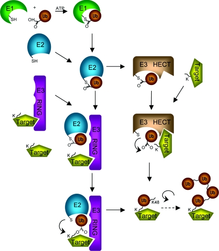
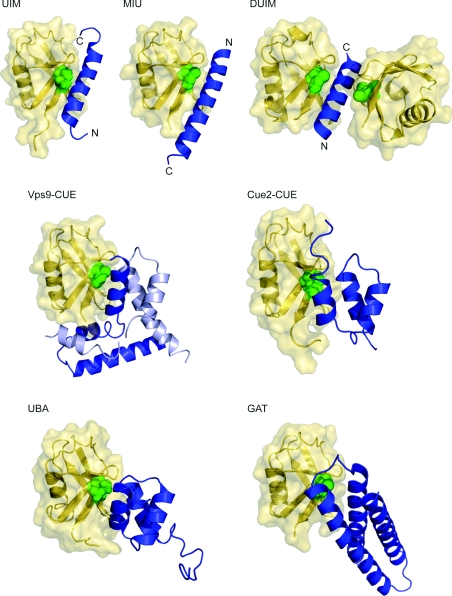
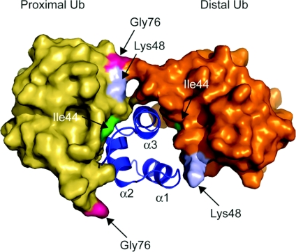
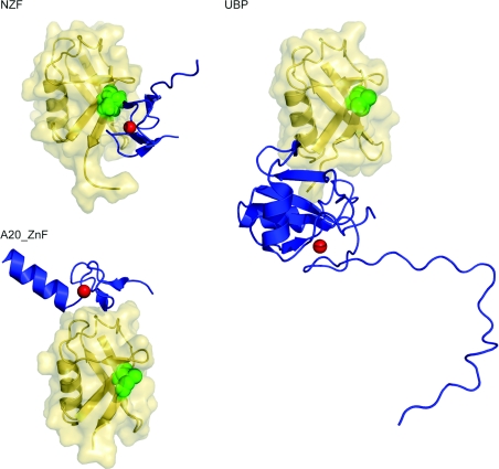
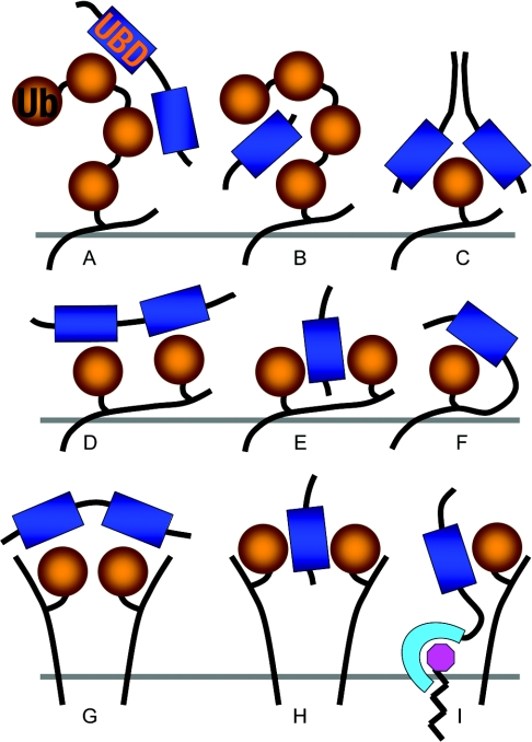
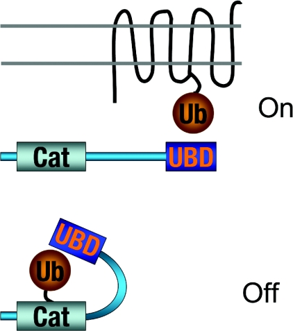
The covalent modification of proteins by ubiquitination is a major regulatory mechanism of protein degradation and quality control, endocytosis, vesicular trafficking, cell-cycle control, stress response, DNA repair, growth-factor signalling, transcription, gene silencing and other areas of biology. A class of specific ubiquitin-binding domains mediates most of the effects of protein ubiquitination. The known membership of this group has expanded rapidly and now includes at least sixteen domains: UBA, UIM, MIU, DUIM, CUE, GAT, NZF, A20 ZnF, UBP ZnF, UBZ, Ubc, UEV, UBM, GLUE, Jab1/MPN and PFU. The structures of many of the complexes with mono-ubiquitin have been determined, revealing interactions with multiple surfaces on ubiquitin. Inroads into understanding polyubiquitin specificity have been made for two UBA domains, whose structures have been characterized in complex with Lys48-linked di-ubiquitin. Several ubiquitin-binding domains, including the UIM, CUE and A20 ZnF (zinc finger) domains, promote auto-ubiquitination, which regulates the activity of proteins that contain them. At least one of these domains, the A20 ZnF, acts as a ubiquitin ligase by recruiting a ubiquitin-ubiquitin-conjugating enzyme thiolester adduct in a process that depends on the ubiquitin-binding activity of the A20 ZnF. The affinities of the mono-ubiquitin-binding interactions of these domains span a wide range, but are most commonly weak, with Kd>100 microM. The weak interactions between individual domains and mono-ubiquitin are leveraged into physiologically relevant high-affinity interactions via several mechanisms: ubiquitin polymerization, modification multiplicity, oligomerization of ubiquitinated proteins and binding domain proteins, tandem-binding domains, binding domains with multiple ubiquitin-binding sites and co-operativity between ubiquitin binding and binding through other domains to phospholipids and small G-proteins.


Figures

Figure 1
Figure 1. Structural features of ubiquitin
(A) Ribbon and surface representations of ubiquitin (Protein data bank identication code: 1UBQ). The C-terminal Gly76 is marked. (B) Location of lysine residues (blue) on ubiquitin. The ubiquitin molecule is shown as surface representation. (C) Major recognition patches on ubiquitin. The hydrophobic patch centred on Ile44 (green), the polar patch centred on Asp58 (blue) and the diglycine patch near the C-terminal Gly76 (pink) are shown.
Figure 2
Figure 2. Major enzymatic pathways of protein ubiquitination
HECT, homologous to E6AP C-terminus; K, lysine; RING, really interesting new gene; Ub, ubiquitin.
Figure 3
Figure 3. Helical ubiquitin-binding domain structures
Ubiquitin molecule (yellow) in ribbon and surface representations is shown with corresponding helical domain (blue) in ribbon representation. Ile44, the centre of the hydrophobic recognition patch on the ubiquitin, is shown as green spheres. Ubiquitin molecules are placed in the same orientation as in Figure 1 for comparison. For the UIM, MIU and DUIM structures, both N- and C-termini are marked. Protein data bank identication codes used are as follows: UIM, 1Q0W; MIU, 2FIF; DUIM, 2D3G; Vps9 CUE, 1P3Q; Cue2-CUE, 1OTR; UBA, 1WR1; GAT, 1YD8. Vps9 CUE domain forms a domain-swapped dimer, shown in blue and light blue. The missing part in Vps9 CUE was modelled on the basis of the apo structure.
Figure 4
Figure 4. A model for polyubiquitin recognition by a UBA domain
The UBA2 domain of hHR23A (blue) with two ubiquitin molecules (yellow and gold) covalently linked via an iso-peptide bond between Lys48 (light blue) of proximal ubiquitin and Gly76 (pink) of distal ubiquitin is shown. The proximal ubiquitin is placed in the same orientation as in Figure 1 for comparison. Ile44 (green) is also indicated. Each helix in the UBA2 domain is labelled. The protein data bank identication code used is 1ZO6.
Figure 5
Figure 5. ZnF domain structures
Three ZnF domains (NZF, UBP and A20 ZnF) are shown (blue) in ribbon representation, with ubiquitin (yellow) in ribbon and surface representations. Ile44, the centre of the hydrophobic recognition patch on the ubiquitin, is shown as green spheres. Ubiquitin molecules are placed in the same orientation as in Figure 1 for comparison. Zinc ions are depicted as red spheres. Protein data bank identication codes used are: A20 ZnF, 2FIF; NZF, 1Q5W; UBP, 2G45.
Figure 6
Figure 6. Ubc-related domain structures
Upper panel: UEV domains of Vps23 and Mms2 are shown (blue) in ribbon representation with ubiquitin (yellow) in ribbon and surface representations. Ile44, the centre of the hydrophobic recognition patch on the ubiquitin, is shown as green spheres. Ubiquitin molecules are placed in the same orientation as in Figure 1 for comparison. Protein data bank identication codes used are: Vps23 UEV, 1UZX; Mms2 UEV, 1ZGU. Lower panels: Ubc5 (blue) is shown on the left with ubiquitin (yellow). The catalytically important Cys85 is depicted as yellow sphere. Yeast Ubc4 is presented in the middle panel for comparison. Again, catalytic Cys86 is shown as yellow sphere. On the right, Ubc9 (blue) complexed with SUMO (small ubiquitin-related modifier) (yellow) is shown [109], as there is no equivalent structure of a Ubc–ubiquitin conjugate available. Leu65 on SUMO, equivalent to Ile44 from sequence alignment, is indicated as green sphere and Cys93 of Ubc9 as yellow sphere. Protein data bank identication codes used are: Ubc5, 2FUH; Ubc4, 1QCQ; Ubc9/SUMO, 1Z5S. The SUMO molecule is also placed in the same orientation as the ubiqutin molecule.
Figure 7
Figure 7. Co-operativity between weak mono-ubiquitin–ubiquitin-binding domain interactions
(A) Recognition of polyubiquitin by tandem mono-ubiquitin-binding domains (UBD) [102,103]. (B) Recognition of polyubiquitin by a two different faces of ubiquitin-binding domain [48,55]. (C) High-affinity binding to mono-ubiquitin by a dimeric ubiquitin-binding domain that uses two different faces on each of the two monomers to recognize two different sites on a single ubiquitin moiety [52]. (D) Recognition of a multimono-ubiquitinated protein by tandem mono-ubiquitin-binding domains [100,110]. (E) Recognition of a multimono-ubiquitinated protein by a two-faced ubiquitin-binding domain [32]. (F) The local concentration facilitates intramolecular inhibition by auto-ubiquitination via a univalent ubiquitin–ubiquitin-binding domain interaction [107]. (G) Recognition of clustered mono-ubiquitinated membrane proteins by tandem mono-ubiquitin-binding domains. (H) Recognition of clustered mono-ubiquitinated membrane proteins by a two-faced ubiquitin-binding domain. (I) Co-operative recognition of a mono-ubiquitinated membrane proteins by a protein containing lipid and mono-ubiquitin-binding domains.
Figure 8
Figure 8. Auto-inhibition of ubiquitin-binding domain proteins
A present model for the regulation of ubiquitin-binding-domain-containing proteins by ubiquitination. UBD, ubiquitin-binding domain.

References

    1. Hershko A., Ciechanover A., Varshavsky A. The ubiquitin system. Nat. Med. 2000;6:1073–1081. - PubMed
    1. Pickart C. M. Mechanisms underlying ubiquitination. Annu. Rev. Biochem. 2001;70:503–533. - PubMed
    1. Weissman A. M. Themes and variations on ubiquitylation. Nat. Rev. Mol. Cell Biol. 2001;2:169–178. - PubMed
    1. Hochstrasser M. Evolution and function of ubiquitin-like protein-conjugation systems. Nat. Cell Biol. 2000;2:E153–E157. - PubMed
    1. Miller J., Gordon C. The regulation of proteasome degradation by multi-ubiquitin chain binding proteins. FEBS Lett. 2005;579:3224–3230. - PubMed

Publication types