. 2006 Nov;146(2):278-86.
doi: 10.1111/j.1365-2249.2006.03198.x.

Complement membrane attack is required for endplate damage and clinical disease in passive experimental myasthenia gravis in Lewis rats

Affiliations

Complement membrane attack is required for endplate damage and clinical disease in passive experimental myasthenia gravis in Lewis rats

J Chamberlain-Banoub et al. Clin Exp Immunol. 2006 Nov.

Abstract

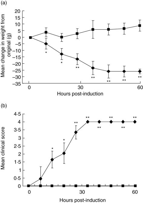
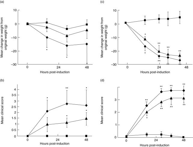
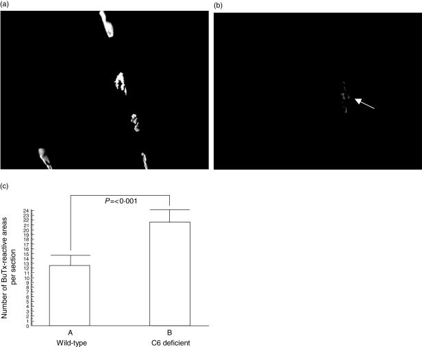
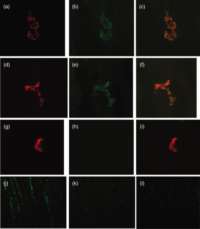
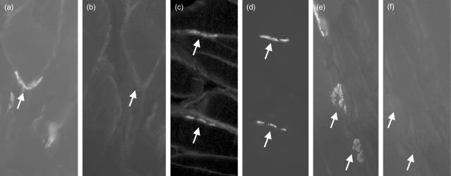
Myasthenia gravis (MG) is a debilitating and potentially fatal neuromuscular disease characterized by the generation of autoantibodies reactive with nicotinic acetylcholine receptors (AChR) that cause loss of AChR from the neuromuscular endplate with resultant failure of neuromuscular transmission. A role for complement (C) in the pathology of human MG has been suggested based upon identification of C activation products in plasma and deposited at the endplate in MG. In the rat model, experimental autoimmune MG (EAMG), C depletion or inhibition restricts clinical disease, further implicating C in pathology. The mechanisms by which C activation drives pathology in MG and EAMG are unclear. Here we provide further evidence implicating C and specifically the membrane attack complex (MAC) in the Lewis rat passive EAMG model of MG. Rats deficient in C6, an essential component of the MAC, were resistant to disease induction and endplate destruction was reduced markedly compared to C6-sufficient controls. After reconstitution with C6, disease severity and endplate destruction in the C6-deficient rats was equivalent to that in controls. The data confirm the essential role of the MAC in the destruction of the endplate in EAMG and raise the prospect of specific MAC inhibition as an alternative therapy in MG patients resistant to conventional treatments.


Figures

Fig. 1
Fig. 1
C6 deficiency protects from clinical disease in the passive experimental autoimmune MG (EAMG) model. Age- and weight-matched groups of C6-deficient (n = 5; squares) and wild-type (n = 6; diamonds) female rats were administered anti-acetylcholine receptor (AChR) at time 0. Weight at time 0 was 230 ± 5 g for the C6-deficient rats (mean ± s.d.) and 234 ± 6·5 g (mean ± s.d.) for wild-type controls. Animals were assessed for change in weight (a) and clinical score (b) as described in Methods. Bars represent ± 1 s.d. Asterisks indicate significant differences between the groups (*P < 0·05; **P < 0·01).
Fig. 2
Fig. 2
Administration of human C6 renders C6-deficient rats susceptible to disease. In two separate experiments, age- and weight-matched groups of six wild-type and C6-deficient female Lewis rats were administered anti-acetylcholine receptor (AChR). In each experiment, wild-type (diamonds), C6-deficient rats without supplementary C6 (squares) and C6-deficient rats with supplementary C6 (triangles) were compared. Animals were assessed for weight change (a,c) and clinical score (b,d) as described in Methods. In experiment 1 (a,b), a single dose (8 mg/kg) of human C6 was given at t = 0; in experiment 2 (c,d), two doses (10 mg/kg each) of humans C6 were given at t = 0 and t = 12 h. Bars represent ± 1 s.d. Where error bars overlapped, only unidirectional bars are shown for clarity. Asterisks indicate significant differences between experimental and control groups (*P < 0·05; **P < 0·01).
Fig. 3
Fig. 3
Loss of acetylcholine receptor (AChR) following experimental autoimmune MG (EAMG) induction requires C6. Soleus muscles were harvested from each rat, frozen sections cut and stained with α-bungarotoxin–rhodamine to identify AChR. Plates show representative fields from C6-deficient (a) and wild-type (b) rats. Using OpenLab imaging software, intensity limits were set, corresponding to positively stained AChR, and the image was density-sliced. The number of discrete areas was measured automatically on the density-sliced image. Twenty fields from each rat were analysed as described in Methods and the mean number of discrete areas was obtained for each rat (c). Columns represent averages for the wild-type (n = 6) and C6-deficient (n = 5) rats and bars show standard deviation for each group. The significance of the difference between groups is shown.
Fig. 4
Fig. 4
Assessment of C3 deposition at the acetylcholine receptor (AChR) and muscle inflammation in wild-type, C6-deficient and naive Lewis rats. Soleus muscle from wild-type (a–c) and C6-deficient (d–f) Lewis rats induced for experimental autoimmune MG (EAMG) and a naive rat (g–i) was harvested and flash-frozen in isopentane as described. Ten μm thin sections were cut and stained for AChR (a, d, g) using rhodamine-conjugated α-bungarotoxin (red staining). Sections were double-stained for activated C3 (C3c; b, e, h) using goat anti-rat C3c, and detected with donkey anti-goat-Ig-fluorescein isothiocyanate (FITC) conjugate (green staining). Sections were mounted in VectorShield and analysed under an inverted fluorescent microscope. Images were merged for detection of co-localization (orange staining; c, f, i). Images were taken at 400× magnification and magnified a further twofold electronically. For assessment of inflammatory cell infiltration, frozen soleus muscle sections from wild-type (j) and C6-deficient (k) Lewis rats induced for EAMG and a naive rat (l) were stained with mouse anti-rat CD68 (ED1) to identify macrophages within the muscle. Images were taken at 200× magnification
Fig. 5
Fig. 5
Assessment of C9/membrane attack complex (MAC) deposition at the acetylcholine receptor (AChR) in wild-type, C6-deficient and naive Lewis rats. Soleus muscle from naive rats (a,b) and wild-type (c,d) or C6-deficient (e,f) Lewis rats induced for experimental autoimmune MG (EAMG) were harvested, and flash-frozen in isopentane as described. Ten μm thin sections were cut and stained for AChR (a,c,e) using rhodamine-conjugated bungarotoxin. Sections were double-stained for rat C9/MAC (b,d,f) using rabbit anti-rat C9, and detected with anti-rabbit-Ig-fluorescein isothiocyanate (FITC) conjugate. Sections were mounted in VectorShield and analysed under an inverted fluorescent microscope. Images were taken at 800× magnification. Arrows in paired images show identical regions stained in one or both.

References

    1. Hughes BW, De Casillas MLM, Kaminski HJ. Pathophysiology of myasthenia gravis. Semin Neurol. 2004;24:21–30. - PubMed
    1. Lindstrom J. Acetylcholine receptors and myasthenia. Muscle Nerve. 2000;23:453–77. - PubMed
    1. Lindstrom J. Nicotinic acetylcholine receptors of muscles and nerves. Comparison of their structures, functional roles and vulnerability to pathology. Ann NY Acad Sci. 2003;998:41–52. - PubMed
    1. Newsom-Davis J, Pinching AJ, Vincent A, Wilson SG. Function of circulating antibody to acetylcholine receptor in myasthenia gravis: investigation by plasma exchange. Neurology. 1978;28:266–72. - PubMed
    1. Nastuk WL, Plescia OJ, Osserman KE. Changes in serum complement activity in patients with myasthenia gravis. Proc Soc Exp Biol Med. 1960;105:177–84. - PubMed

Publication types

MeSH terms