. 2006 Oct 30;580(25):5885-93.
doi: 10.1016/j.febslet.2006.09.048. Epub 2006 Oct 2.

A novel analytical method for in vivo phosphate tracking

Affiliations

A novel analytical method for in vivo phosphate tracking

Hong Gu et al. FEBS Lett. .

Erratum in

  • FEBS Lett. 2007 Feb 6;581(3):579

Abstract

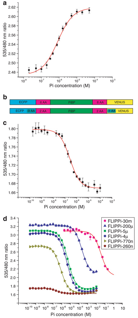
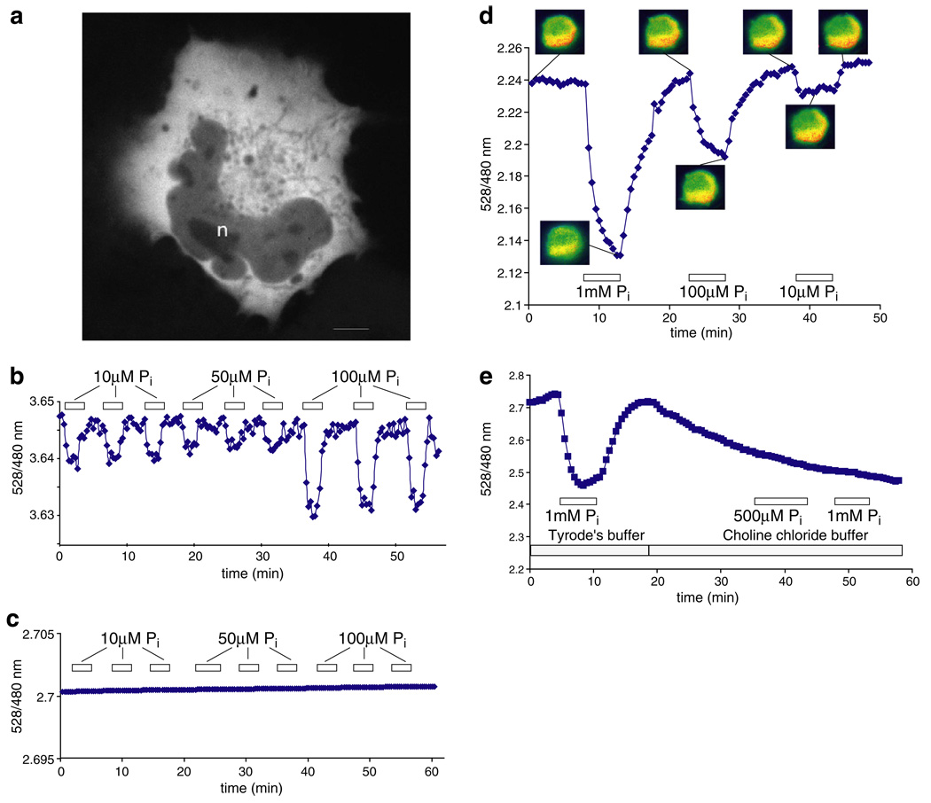
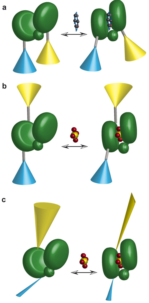
Genetically-encoded fluorescence resonance energy transfer (FRET) sensors for phosphate (P(i)) (FLIPPi) were engineered by fusing a predicted Synechococcus phosphate-binding protein (PiBP) to eCFP and Venus. Purified fluorescent indicator protein for inorganic phosphate (FLIPPi), in which the fluorophores are attached to the same PiBP lobe, shows P(i)-dependent increases in FRET efficiency. FLIPPi affinity mutants cover P(i) changes over eight orders of magnitude. COS-7 cells co-expressing a low-affinity FLIPPi and a Na(+)/P(i) co-transporter exhibited FRET changes when perfused with 100 microM P(i), demonstrating concentrative P(i) uptake by PiT2. FLIPPi sensors are suitable for real-time monitoring of P(i) metabolism in living cells, providing a new tool for fluxomics, analysis of pathophysiology or changes of P(i) during cell migration.


Figures

Fig. 1
Fig. 1
(a) In vitro ligand-dependent FRET changes of FLIPPi-WT purified nanosensors. Non-linear regression best fit and error bars are shown. (b) FLIPPi-WT construct (above); FLIPPi-260n construct (below; nine amino acids were deleted from the C-terminus of the eCFP, one from the N-terminus of Venus, and four from each binding protein-fluorophore linker). (c) and (d) In vitro ligand-dependent FRET changes of FLIPPi-260n (c) and affinity mutants of the FLIPPi-260n construct (d; Table 1).
Fig. 2
Fig. 2
The predicted structure of PiBP in alignment with the E. coli PiBP (PDB 1a40). (a) Synechococcus PiBP structural model, predicted by 3D-JIGSAW. Phosphate is in yellow, and the N- and C-termini in red. Terminal lobe, blue; other lobe, green. (b) Alignment of the model (blue) with the crystal structure of E. coli (green). (c) Alignment of binding sites. The only sequence difference is Glu70 in FLIPPi and Asp56 in E. coli. The Synechococcus structure has likely been poorly modeled in the region of Glu70 and Ser161 (phosphate was not present in the 3D-JIGSAW modeling step). Green: E. coli; blue: FLIPPi; yellow: phosphate.
Fig. 3
Fig. 3
Specificity and pH sensitivity of FLIPPi-5µ. (a) Selectivity of FLIPPi-5µ for phosphate over nitrate and sulfate. Solid lines were plotted by curve fitting. (b) Phosphate binding of FLIPPi-5µ was tested in 20 mM Tris buffers adjusted to pH 6.8, 7.0, 7.2, 7.4 and 7.6, then titrated with phosphate solutions of the same respective pH. Statistical analysis (ANOVA) showed no significant difference regarding the maximal ratio change between pH 6.8 and 7.4.
Fig. 4
Fig. 4
FRET ratio change in response to Pi perfusion of Pi-starved CHO cells, and COS-7 cells co-expressing a Na+/Pi cotransporter. (a) Confocal image of a COS-7 cell expressing FLIPPi-30m. Fluorescence is largely excluded from the nucleus (n). The scale bar represents 5 µm. (b) The low-affinity “working” sensor FLIPPi-30m in Pi-starved CHO cells. (c) The high-affinity “control” sensor FLIPPi-5µ is saturated and does not respond to Pi perfusion. (d) The FLIPPi-30m sensor in resting COS-7 cells co-expressing the sodium-dependent phosphate cotransporter PiT2. (e) In sodium-free modified Tyrode’s choline buffer, the response of the sensor to Pi is abolished. The boxed numbers indicate the Pi concentration used for perfusion.
Fig. 5
Fig. 5
Cartoon models of FLIPPi sensor function [20]. (a) Model of FLIP sensor developed from type I periplasmic binding proteins (e.g. FLIPglu glucose binding protein [14,15]). Fluorophores (eCFP, cyan; Venus, yellow) attached on two different lobes of a binding protein (consisting of two lobes connected by a hinge, green) via linkers (grey rods). Ligand binding transduces a hinge-binding motion propagated through a lever arm to macroscopically reorient the relative excitation–emission dipole. (b) Model of a FLIP sensor developed from type II PiBPs, in which both fluorophores are attached to the same lobe. Allosteric effects of domain closure propagate into relative dipole reorientation. (c) Shortening or rigidification of a binding protein–fluorophore linker increases the degree of allosteric coupling between ligand binding and fluorophore reorientation. One possible scenario is shown here, in which the closure of the PiBP leads to a restriction in the rotational freedom of eYFP, illustrated as a change in the cone diameter.

References

    1. Collins JF, Bai L, Ghishan FK. The SLC20 family of proteins: dual functions as sodium-phosphate cotransporters and viral receptors. Pflügers Arch. 2004;447:647–652. - PubMed
    1. Vidal G, Gallis JL, Dufour S, Canioni P. NMR studies of inorganic phosphate compartmentation in the isolated rat liver during acidic perfusion. Arch. Biochem. Biophys. 1997;337:317–325. - PubMed
    1. Wang ZM, Choudhary A, Ledvina PS, Quiocho FA. Fine-tuning the specificity of the periplasmic phosphate transport receptor – site-directed mutagenesis, ligand-binding, and crystallographic studies. J. Biol. Chem. 1994;269:25091–25094. - PubMed
    1. Medveczky N, Rosenberg H. Phosphate-binding protein of Escherichia coli. Biochim. Biophys. Acta. 1970;211:158.
    1. Brune M, Hunter JL, Corrie JET, Webb MR. Direct, real-time measurement of rapid inorganic-phosphate release using a novel fluorescent probe and its application to actomyosin subfragment-1 ATPase. Biochemistry. 1994;33:8262–8271. - PubMed

Publication types

MeSH terms