. 2007 Jan 10;176(1):94-108.
doi: 10.1016/j.bbr.2006.08.026. Epub 2006 Oct 10.

Modeling early cortical serotonergic deficits in autism

Affiliations

Modeling early cortical serotonergic deficits in autism

Carolyn B Boylan et al. Behav Brain Res. .

Abstract

Autism is a developmental brain disorder characterized by deficits in social interaction, language and behavior. Brain imaging studies demonstrate increased cerebral cortical volumes and micro- and macro-scopic neuroanatomic changes in children with this disorder. Alterations in forebrain serotonergic function may underlie the neuroanatomic and behavioral features of autism. Serotonin is involved in neuronal growth and plasticity and these actions are likely mediated via serotonergic and glutamatergic receptors. Few animal models of autism have been described that replicate both etiology and pathophysiology. We report here on a selective serotonin (5-HT) depletion model of this disorder in neonatal mice that mimics neurochemical and structural changes in cortex and, in addition, displays a behavioral phenotype consistent with autism. Newborn male and female mice were depleted of forebrain 5-HT with injections of the serotonergic neurotoxin, 5,7-dihydroxytryptamine (5,7-DHT), into the bilateral medial forebrain bundle (mfb). Behavioral testing of these animals as adults revealed alterations in social, sensory and stereotypic behaviors. Lesioned mice showed significantly increased cortical width. Serotonin immunocytochemistry showed a dramatic long-lasting depletion of 5-HT containing fibers in cerebral cortex until postnatal day (PND) 60. Autoradiographic binding to high affinity 5-HT transporters was significantly but transiently reduced in cerebral cortex of 5,7-DHT-depleted mice. AMPA glutamate receptor binding was decreased at PND 15. We hypothesize that increased cerebral cortical volume and sensorimotor, cognitive and social deficits observed in both 5-HT-depleted animals and in individuals with autism, may be the result of deficiencies in timely axonal pruning to key cerebral cortical areas.


Figures

Figure 1
Figure 1. PND 7 age normal control
This figure illustrates the pattern of 5-HT immunostaining in a parasagittal section. Overlayed are labels of the different brain regions that the raphe serotonergic cells projects to: frontal cortex (fr ctx), parietal cortex (par ctx), occipital cortex (occ ctx), hippocampus (hippo), striatum, thalamus, midbrain and cerebellum (cblm). Although not present in this section, the approximate locations of the dorsal raphe and the medial forebrain bundle (mfb) are demarcated. Arrows illustrate the path the serotonergic axons travel from the dorsal raphe through the mfb to the cortex and hippocampus.
Figure 2
Figure 2. Effects of the 5,7-DHT lesion on serotonin immunocytochemistry
Bilateral lesions to the mfb with 5,7-DHT on the day of birth produced near complete loss of 5-HT immunoreactivity in cerebral cortex and hippocampus at PND 7, 15 and 30 but did not affect the distribution of these fibers in brain regions unaffected by lesion. An arrow points to the remaining 5-HT staining in layer IV of barrel field cortex in lesioned mice at PND 7. This is the site where thalamocortical axons transiently take up serotonin at this age.
Figure 3
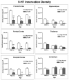
Figure 3. Density of 5-HT containing fibers
Densitometric analysis of 5-HT immunoreactivity showed a decreased density of 5-HT containing fibers in most regions of cerebral cortex and hippocampus in 5,7-DHT-treated mice at most of the ages studied (*p<0.05, **p<0.01). In contrast, the density of 5-HT axons was unchanged in thalamus and cerebellum after forebrain 5-HT depletion.
Figure 4
Figure 4. Localization of AMPA receptor sites
Representative autoradiograms of AMPA receptor binding in coronal sections from age normal and 5,7-DHT-depleted PND 7, 15 and 30 mice. Of the few AMPA sites present at PND 7, most were in the hippocampus, striatum and ventral cortex. By PND15, the density of AMPA receptors increased markedly in dorsal cortex, striatum and hippocampus. A similar pattern of AMPA recptors was present at PND 30. The look up table in the PND 15 lesioned autoradiogram indicates that cooler colors refer to lower densities of receptors and warmer colors for higher densities of receptors.
Figure 5
Figure 5. Localization of NMDA receptor sites
Representative autoradiograms of NMDA receptor binding in coronal sections from age normal and 5,7-DHT-depleted PND 7, 15 and 30 mice. At PND 7very few NMDA sites present. By PND 15, NMDA receptor density increased in the cortex and hippocampus. With age, the density of these receptors increased throughout the cerebral cortex for all groups of mice.
Figure 6
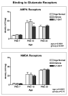
Figure 6. Density of AMPA and NMDA receptor binding
The mean density ± s.e.m. of AMPA and NMDA receptors in cerebral cortex from PND 7, 15, and 30 age normal control, vehicle-injected, and 5,7-DHT-lesioned mice. AMPA receptor binding density in cerebral cortex of 5,7-DHT-lesioned mice is significantly less compared to age normal and vehicle-injected animals at PND 15 (p<0.05) but was unchanged at PND 7 and 30. NMDA receptor binding density was unchanged at PND 7, 15 or 30. A two way ANOVA showed that the density of both AMPA and NMDA receptors increased with age (p<0.0001). This analysis also showed a trend for a group effect for AMPA (p=0.057) and NMDA (p=0.103) receptors.
Figure 7
Figure 7. Social Transmission of Food Preference Task
The amount of cued (demonstrated) flavor consumed by neonatally 5,7-DHT-lesioned males and females was significantly lower (p=0.038; *p<0.05) than in age normal control mice. Vehicle-injected mice were also tested and consumed quantities of cued food exceeding the consumption of age normal controls. Non-cued food consumption in all groups was below the measurable scale for this graph. Serotonin-depleted mice of either sex consumed approximately 50% as much non-cued as cued food. These differences were not statistically significant. However, age normal and vehicle-injected control mice consumed less non-cued food as compared with cued food (p=0.05).
Figure 8
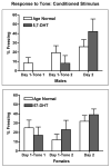
Figure 8. CCFC sound
None of the male 5,7-DHT males responded with freezing bouts to the first exposure of a tone cue, while all age matched normal controls did. Upon pairing the sound cue with foot-shock later on Day 1, the lesioned mice did respond with freezing and 24 hours later, male lesioned mice responded with increased freezing to re-exposure to the sound cue (differences on Day 1 tone 2 were not significant, differences on Day 2 approached significance). No significant differences or trends were observed in the females.
Figure 9
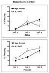
Figure 9. CCFC context
Female mice, in general, showed more freezing bouts then males. All females and male age normal control mice increased the freezing response when re-exposed to the original conditioning chamber on Day 3 (compared to Day 2) and freezing responses in the cued environment significantly increased from first exposure to the conditioned stimulus on Day 1 (p=0.0017 for male age matched controls, p=0.004 and 0.007 respectively for female age matched controls and female lesioned mice). In contrast, 5,7-DHT lesioned males actually decreased their freezing response on Day 3 compared to Day 2, and had markedly lower freezing responses compared to age normal control males and showed no significant differences in freezing between first exposure on Day 1 and Day 3.

References

    1. Akshoomoff N, Lord C, Lincoln AJ, Courchesne RY, Carper RA, Townsend J, Courchesne E. Outcome classification of preschool children with autism spectrum disorders using MRI brain measures. J Am Acad Child Adolesc Psychiatry. 2004;43:349–357. - PubMed
    1. Anderson GM, Horne WC, Chatterjee D, Cohen DJ. The hyperserotonemia of autism. Ann N Y Acad Sci. 1990;600:331–340. discussion 341–332. - PubMed
    1. Aylward EH, Minshew NJ, Field K, Sparks BF, Singh N. Effects of age on brain volume and head circumference in autism. Neurology. 2002;59:175–183. - PubMed
    1. Bailey A, Luthert P, Dean A, Harding B, Janota I, Montgomery M, Rutter M, Lantos P. A clinicopathological study of autism. Brain. 1998;121 ( Pt 5):889–905. - PubMed
    1. Baranek GT. Autism during infancy: a retrospective video analysis of sensory-motor and social behaviors at 9–12 months of age. J Autism Dev Disord. 1999;29:213–224. - PubMed

Publication types

MeSH terms