. 2007 Jan;81(1):319-31.
doi: 10.1128/JVI.01842-06. Epub 2006 Oct 11.

Cytoplasmic residues of herpes simplex virus glycoprotein gE required for secondary envelopment and binding of tegument proteins VP22 and UL11 to gE and gD

Affiliations

Cytoplasmic residues of herpes simplex virus glycoprotein gE required for secondary envelopment and binding of tegument proteins VP22 and UL11 to gE and gD

Aaron Farnsworth et al. J Virol. 2007 Jan.

Abstract

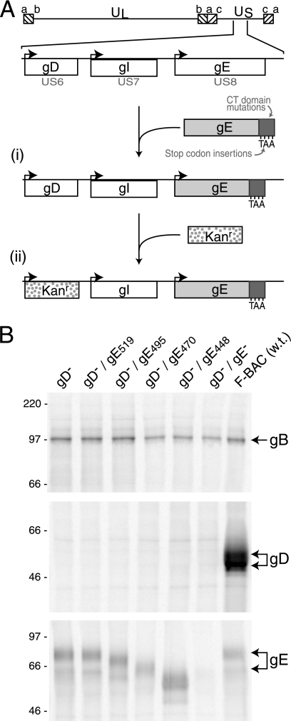
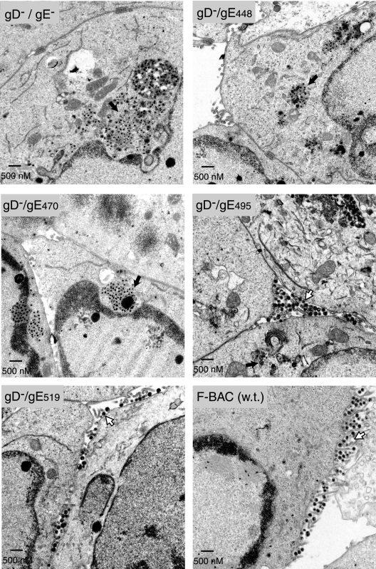
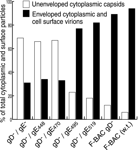
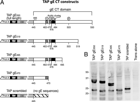
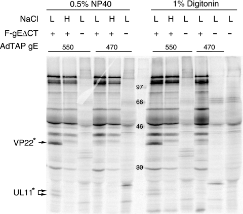
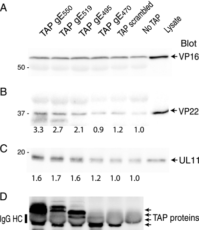
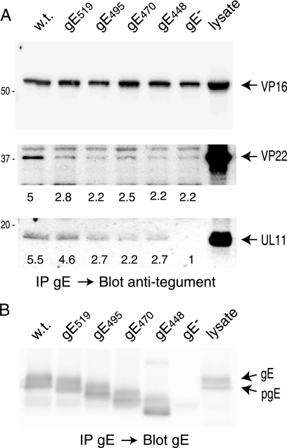
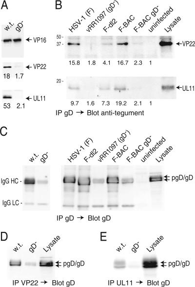
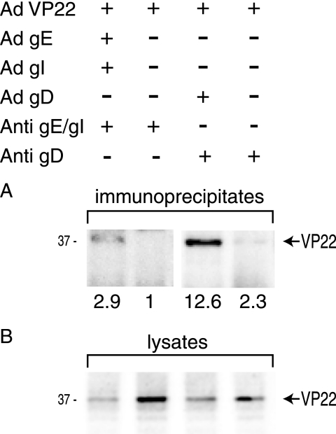
The final assembly of herpes simplex virus (HSV) involves binding of tegument-coated capsids to viral glycoprotein-enriched regions of the trans-Golgi network (TGN) as enveloped virions bud into TGN membranes. We previously demonstrated that HSV glycoproteins gE/gI and gD, acting in a redundant fashion, are essential for this secondary envelopment. To define regions of the cytoplasmic (CT) domain of gE required for secondary envelopment, HSVs lacking gD and expressing truncated gE molecules were constructed. A central region (amino acids 470 to 495) of the gE CT domain was important for secondary envelopment, although more C-terminal residues also contributed. Tandem affinity purification (TAP) proteins including fragments of the gE CT domain were used to identify tegument proteins VP22 and UL11 as binding partners, and gE CT residues 470 to 495 were important in this binding. VP22 and UL11 were precipitated from HSV-infected cells in conjunction with full-length gE and gE molecules with more-C-terminal residues of the CT domain. gD also bound VP22 and UL11. Expression of VP22 and gD or gE/gI in cells by use of adenovirus (Ad) vectors provided evidence that other viral proteins were not necessary for tegument/glycoprotein interactions. Substantial quantities of VP22 and UL11 bound nonspecifically onto or were precipitated with gE and gD molecules lacking all CT sequences, something that is very unlikely in vivo. VP16 was precipitated equally whether gE/gI or gD was present in extracts or not. These observations illustrated important properties of tegument proteins. VP22, UL11, and VP16 are highly prone to binding nonspecifically to other proteins, and this did not represent insolubility during our assays. Rather, it likely reflects an inherent "stickiness" related to the formation of tegument. Nevertheless, assays involving TAP proteins and viral proteins expressed by HSV and Ad vectors supported the conclusion that VP22 and UL11 interact specifically with the CT domains of gD and gE.


Figures

FIG. 1.
FIG. 1.
Construction of recombinant viruses. (A) The genome of HSV-1, including the US6 (gD), US7 (gI), and US8 (gE) genes, is depicted (44). (i) The insertion of stop codons in the gE CT domain after residues 448, 470, 495, and 519 was previously described (23). (ii) A kanamycin resistance gene cassette was inserted into the coding sequences of gD, and this was transferred into BACs containing truncated versions of gE. (B) Vero cells were infected with F-BAC gD, F-BAC gD/gE519, F-BAC gD/gE495, F-BAC gD/gE470, F-BAC gD/gE448, F-BAC gD/gE, or wild-type F-BAC HSV-1 (w.t.). The cells were labeled with [35S]methionine-cysteine, and gB, gD, or gE was immunoprecipitated from cell extracts by using MAb 15βB2, DL6, or 3114, respectively. The positions of gB, gD, and gE are indicated on the right side of the panel.
FIG. 2.
FIG. 2.
Electron micrographs of cells infected with F-BAC gD/gE CT mutants. Human HEC-1A epithelial cells were infected with F-BAC gD/gE, F-BAC gD/gE448, F-BAC gD/gE470, F-BAC gD/gE495, F-BAC gD/gE519, or F-BAC (w.t.) for 16 h. The cells were fixed and processed for electron microscopy. Black arrows point to aggregates of unenveloped capsids, while white arrows point to enveloped virions on the surfaces of cells.
FIG. 3.
FIG. 3.
Distributions of virus particles in cells infected by gD/gE CT mutants. Randomly selected sections of HSV-infected HEC-1A cells were characterized by electron microscopy, and unenveloped nucleocapsids in the nucleus and cytoplasm and enveloped virions in the perinuclear space, in the cytoplasm, and on the cell surface were counted. In the figure, values for unenveloped capsids in the cytoplasm (white bars) are compared to those for enveloped virions in the cytoplasm combined with virions on cell surfaces (black bars).
FIG. 4.
FIG. 4.
Construction of TAP-gE fusion proteins. (A) The TAP domain was composed of two tandem protein A (IgG-binding) domains separated from a CBD by a tobacco etch virus protease cleavage site. The TAP domain was fused N-terminal of the entire gE CT domain, beginning with three arginine residues that are adjacent to the gE transmembrane domain. Other TAP fusion proteins included truncated versions of the gE CT domain: gE519, gE495, and gE470. Also shown is a construct designated TAP scrambled, which contains 25 random amino acids unrelated to the gE CT domain. (B) Vero cells were coinfected with Ad vectors expressing TAP/gE550, TAP/gE519, TAP/gE495, TAP/gE470, or TAP scrambled and Adtet-trans or with Adtet-trans alone. Cell extracts were subjected to electrophoresis on polyacrylamide gels, and then proteins were transferred to the PVDF membrane and probed with an anti-CBD rabbit antibody. Molecular mass markers of 50, 37, 25, and 20 kDa are indicated.
FIG. 5.
FIG. 5.
Interactions between TAP/gE fusion proteins and HSV proteins. Human HaCaT keratinocytes were infected with Ad expressing either TAP/gE550 or TAP/gE470 for 18 h and then infected with F-gEΔCT (a gE-null mutant) for an additional 7 to 8 h. The cells were radiolabeled with [35S]methionine-cysteine for 3 h, and then cell extracts were made using 0.5% NP-40 lysis buffer or 1% digitonin lysis buffer and low salt (L) (100 mM NaCl) or high salt (H) (500 mM NaCl). Extracts were centrifuged at 60,000 × g and supernatants incubated with IgG-Sepharose at 4°C. Proteins were eluted and subjected to electrophoresis. Two sets of bands, designated VP22* and UL11*, were observed when cells were infected with TAP/gE550 and F-gEΔCT but not with TAP/gE470 and F-gEΔCT and not without F-gEΔCT. Molecular mass markers of 97, 66, 46 and 30 kDa are indicated.
FIG. 6.
FIG. 6.
Coimmunoprecipitation of VP16, VP22, or UL11 with TAP/gE fusion proteins. HaCaT cells were coinfected with Ad vectors expressing TAP/gE550, TAP/gE519, TAP/gE495, TAP/gE470, TAP scrambled, and AdTet trans or were not infected with an Ad vector (No TAP) for 18 h. The cells were subsequently infected with HSV F-gEΔCT and harvested 12 h later in 0.5% NP-40 lysis buffer. Cell extracts were incubated with IgG-Sepharose and washed, and proteins were eluted and subjected to electrophoresis before transfer to PVDF membranes. Membranes were probed with rabbit polyclonal antibodies specific for VP16 (A), VP22 (B), UL11 (C), or anti-protein A antibodies to detect TAP proteins (D). Note that IgG eluted from IgG-Sepharose and the heavy chain comigrated with TAP proteins, as indicated in panel D. VP22 and UL11 levels were quantified using IP Lab Gel software and compared to immunoprecipitation levels for extracts containing no TAP proteins (No TAP), which were set at 1.
FIG. 7.
FIG. 7.
Coimmunoprecipitation of VP16, VP22, or UL11 with gE from HSV-infected cells. HaCaT cells were infected with HSV F-BAC (w.t.) expressing wild-type gE, F-BAC gE519, F-BAC gE495, F-BAC gE470, F-BAC gE448, or F-BAC gE for 12 h. Cell extracts were made using 0.5% NP-40 lysis buffer and gE immunoprecipitated with anti-gE MAb 3114. Precipitated proteins and a sample representing 20% of the cell lysate were subjected to electrophoresis, and proteins were transferred to membranes and then Western blotted with rabbit polyclonal antibodies specific for anti-VP16 (panel A, top), anti-VP22 (panel A, middle), anti-UL11 (panel A, bottom), or gE (B). VP22 and UL11 were quantified using IP Lab Gel software, with the background value (set at 1.0) determined by counting the pixels in the blot regions containing no obvious proteins.
FIG. 8.
FIG. 8.
Coimmunoprecipitation of VP22 and UL11 with gD from HSV-infected cells. (A) HaCaT cells were infected with F-BAC (w.t.) or F-BAC gD for 12 h, and then cell extracts were made using 0.5% NP-40 lysis buffer. gD was immunoprecipitated using MAb DL6, proteins were subjected to electrophoresis and then transferred to membranes, and the membranes were probed with rabbit polyclonal anti-VP16 (upper panel), anti-VP22 (middle panel), or anti-UL11 (lower panel) antibodies. (B) HaCaT cells were infected with wild-type HSV-1 strain F, vRR1097 (a gD-null mutant), F-dl2 (expressing a gD lacking the CT domain), F-BAC, or F-BAC gD for 12 h or were left uninfected. Cell extracts were made using 0.5% NP-40 lysis buffer, and gD was immunoprecipitated using anti-gD MAb DL6. Precipitated proteins and a sample representing 20% of the cell extract (Lysate) were subjected to electrophoresis, transferred to membranes, and probed with anti-VP22 or anti-UL11 rabbit antibodies. The numbers shown in panels A and B were derived as described in the legend to Fig. 7, with the background value (set at 1) corresponding to the blot regions with no obvious proteins. (C) Samples immunoprecipitated as described for panels A and B were also probed with rabbit anti-gD antibodies. The IgG-heavy and -light chains (indicated by HC and LC, respectively) derived from mouse MAb DL6 used to precipitate gD were detected through cross-reaction with secondary antibodies and were most obvious in samples from cells lacking gD. (D and E) VP22 or UL11 was precipitated using rabbit polyclonal antibodies, and samples were blotted with mouse anti-gD MAb DL6. In panel D, some IgG-heavy chains were detected by the secondary antibodies, most obviously in samples from the gD mutant.
FIG. 9.
FIG. 9.
VP22 binds to gE/gI and gD in the absence of other HSV proteins. HaCaT cells were infected for 24 h with nonreplicating Ad vectors expressing VP22, gE/gI, and gD in conjunction with AdTet-trans, as indicated in the upper panel. Cell extracts were made using 0.5% NP-40 lysis buffer. (A) gE/gI immunoprecipitated with pooled gE-specific MAb 3114 and gI-specific MAb 3104 (Anti-gE/gI) or gD precipitated with anti-gD MAb DL6 (Anti-gD), as indicated. Immunoprecipitated proteins were probed using antibodies specific to VP22. (B) Approximately 5% of the cell extract was subjected to electrophoresis, transferred to membranes, and then blotted with anti-VP22 antibodies.

References

    1. Baines, J. D., R. J. Jacob, L. Simmerman, and B. Roizman. 1995. The herpes simplex virus 1 UL11 proteins are associated with cytoplasmic and nuclear membranes and with nuclear bodies of infected cells. J. Virol. 69:825-833. - PMC - PubMed
    1. Baines, J. D., and B. Roizman. 1992. The UL11 gene of herpes simplex virus 1 encodes a function that facilitates nucleocapsid envelopment and egress from cells. J. Virol. 66:5168-5174. - PMC - PubMed
    1. Ball, J. M., Z. Moldoveanu, L. R. Melsen, P. A. Kozlowski, S. Jackson, M. J. Mulligan, J. F. Mestecky, and R. W. Compans. 1995. A polarized human endometrial cell line that binds and transports polymeric IgA. In Vitro Cell. Dev. Biol. Anim. 31:196-206. - PubMed
    1. Beitia Ortiz de Zarate, I., K. Kaelin, and F. Rozenberg. 2004. Effects of mutations in the cytoplasmic domain of herpes simplex virus type 1 glycoprotein B on intracellular transport and infectivity. J. Virol. 78:1540-1551. - PMC - PubMed
    1. Boukamp, P., R. T. Petrussevska, D. Breitkreutz, J. Hornung, A. Markham, and N. E. Fusenig. 1988. Normal keratinization in a spontaneously immortalized aneuploid human keratinocyte cell line. J. Cell Biol. 106:761-771. - PMC - PubMed

Publication types

MeSH terms

Substances

LinkOut - more resources