Construction and mutagenesis of an artificial bicistronic tick-borne encephalitis virus genome reveals an essential function of the second transmembrane region of protein e in flavivirus assembly
- PMID: 17035331
- PMCID: PMC1676298
- DOI: 10.1128/JVI.01540-06
Construction and mutagenesis of an artificial bicistronic tick-borne encephalitis virus genome reveals an essential function of the second transmembrane region of protein e in flavivirus assembly
Abstract
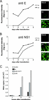
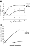
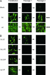
Flaviviruses have a monopartite positive-stranded RNA genome, which serves as the sole mRNA for protein translation. Cap-dependent translation produces a polyprotein precursor that is co- and posttranslationally processed by proteases to yield the final protein products. In this study, using tick-borne encephalitis virus (TBEV), we constructed an artificial bicistronic flavivirus genome (TBEV-bc) in which the capsid protein and the nonstructural proteins were still encoded in the cap cistron but the coding region for the surface proteins prM and E was moved to a separate translation unit under the control of an internal ribosome entry site element inserted into the 3' noncoding region. Mutant TBEV-bc was shown to produce particles that packaged the bicistronic RNA genome and were infectious for BHK-21 cells and mice. Compared to wild-type controls, however, TBEV-bc was less efficient in both RNA replication and infectious particle formation. We took advantage of the separate expression of the E protein in this system to investigate the role in viral assembly of the second transmembrane region of protein E (E-TM2), a second copy of which was retained in the cap cistron to fulfill its other role as an internal signal sequence in the polyprotein. Deletion analysis and replacement of the entire TBEV E-TM2 region with its counterpart from another flavivirus revealed that this element, apart from its role as a signal sequence, is important for virion formation.
Figures


Similar articles
-
Selection and analysis of mutations in an encephalomyocarditis virus internal ribosome entry site that improve the efficiency of a bicistronic flavivirus construct.J Virol. 2007 Nov;81(22):12619-29. doi: 10.1128/JVI.01017-07. Epub 2007 Sep 12. J Virol. 2007. PMID: 17855533 Free PMC article.
-
Steps of the tick-borne encephalitis virus replication cycle that affect neuropathogenesis.Virus Res. 2005 Aug;111(2):161-74. doi: 10.1016/j.virusres.2005.04.007. Virus Res. 2005. PMID: 15871909 Review.
-
Immunization with recombinant vaccinia viruses expressing structural and part of the nonstructural region of tick-borne encephalitis virus cDNA protect mice against lethal encephalitis.J Biotechnol. 1996 Jan 26;44(1-3):97-103. doi: 10.1016/0168-1656(95)00141-7. J Biotechnol. 1996. PMID: 8717392
-
Resuscitating mutations in a furin cleavage-deficient mutant of the flavivirus tick-borne encephalitis virus.J Virol. 2005 Sep;79(18):11813-23. doi: 10.1128/JVI.79.18.11813-11823.2005. J Virol. 2005. PMID: 16140758 Free PMC article.
-
Molecular biology of flaviviruses.Novartis Found Symp. 2006;277:23-39; discussion 40, 71-3, 251-3. Novartis Found Symp. 2006. PMID: 17319152 Review.
Cited by
-
An in vitro recombination-based reverse genetic system for rapid mutagenesis of structural genes of the Japanese encephalitis virus.Virol Sin. 2015 Oct;30(5):354-62. doi: 10.1007/s12250-015-3623-2. Epub 2015 Sep 30. Virol Sin. 2015. PMID: 26463213 Free PMC article.
-
The helical domains of the stem region of dengue virus envelope protein are involved in both virus assembly and entry.J Virol. 2011 May;85(10):5159-71. doi: 10.1128/JVI.02099-10. Epub 2011 Mar 2. J Virol. 2011. PMID: 21367896 Free PMC article.
-
Functional analysis of potential carboxy-terminal cleavage sites of tick-borne encephalitis virus capsid protein.J Virol. 2008 Mar;82(5):2218-29. doi: 10.1128/JVI.02116-07. Epub 2007 Dec 26. J Virol. 2008. PMID: 18160443 Free PMC article.
-
Synergistic Internal Ribosome Entry Site/MicroRNA-Based Approach for Flavivirus Attenuation and Live Vaccine Development.mBio. 2017 Apr 18;8(2):e02326-16. doi: 10.1128/mBio.02326-16. mBio. 2017. PMID: 28420742 Free PMC article.
-
A trans-complementing recombination trap demonstrates a low propensity of flaviviruses for intermolecular recombination.J Virol. 2010 Jan;84(1):599-611. doi: 10.1128/JVI.01063-09. J Virol. 2010. PMID: 19864381 Free PMC article.
References
Publication types
MeSH terms
Substances
LinkOut - more resources
Full Text Sources
Other Literature Sources
Miscellaneous

