

Review
doi: 10.1523/JNEUROSCI.3829-06.2006.
Disease-modifying pathways in neurodegeneration
Affiliations
- PMID: 17035516
- PMCID: PMC6674695
- DOI: 10.1523/JNEUROSCI.3829-06.2006
Item in Clipboard
Review
Disease-modifying pathways in neurodegeneration
J Neurosci.
.
No abstract available
Figures
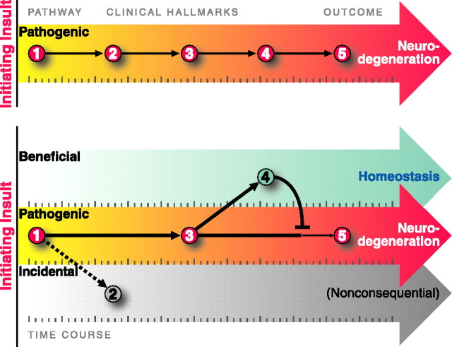
Neurodegenerative diseases display phenotypes that can be beneficial, pathogenic, or incidental. Whether an observed change is pathogenic or beneficial is a common question that has enormous therapeutic importance. The top depicts the time course over which clinical hallmarks appear based on standard pathology studies. However, some changes may actually be beneficial (bottom, 4) or inconsequential (bottom, 2), rather than pathogenic. Activation of homeostatic pathways may mitigate pathogenic perturbations, and promoting these beneficial coping responses could be effective therapies. The timing of the events can further confound recognition of the true disease pathways. If pathogenic processes elicit nearly simultaneous coping responses, they will be highly correlated with each other and with the course of disease. New strategies will enable us to identify disease-modifying pathways and may identify new therapeutic targets for these devastating diseases.
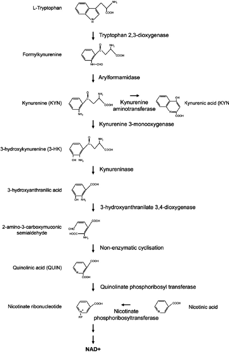
The kynurenine pathway of tryptophan degradation. This metabolic pathway is abnormal in HD patients and transgenic mouse models of HD. Toxic metabolites in this pathway generated by microglia may contribute to neurodegeneration in HD by causing excitotoxicity and/or generation of reactive oxygen species.
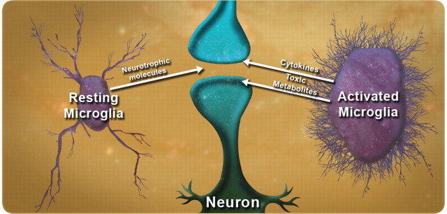
The yin and yang of microglia in neurodegeneration. Under normal conditions, microglia provide trophic support to surrounding neurons. However, in certain disease states, they can become dysfunctional and secrete toxic molecules that may contribute to the demise of surrounding neurons. Such a scenario might be caused by accumulation of misfolded protein directly in microglia, as may occur in HD and ALS.
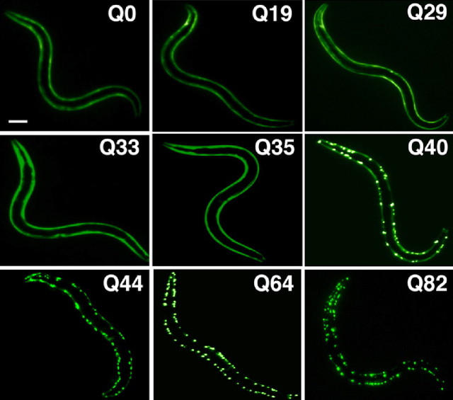
PolyQ length-dependent aggregation in C. elegans. Length-dependent aggregation of polyQ–YFP fusion proteins in C. elegans. Epifluorescence micrographs of 3- to 4-d-old C. elegans expressing different lengths of polyQ–YFP (Q0, Q19, Q29, Q33, Q35, Q40, Q44, Q64, and Q82). Scale bar, 0.1 mm. [Adapted from Morley et al. (2002).].
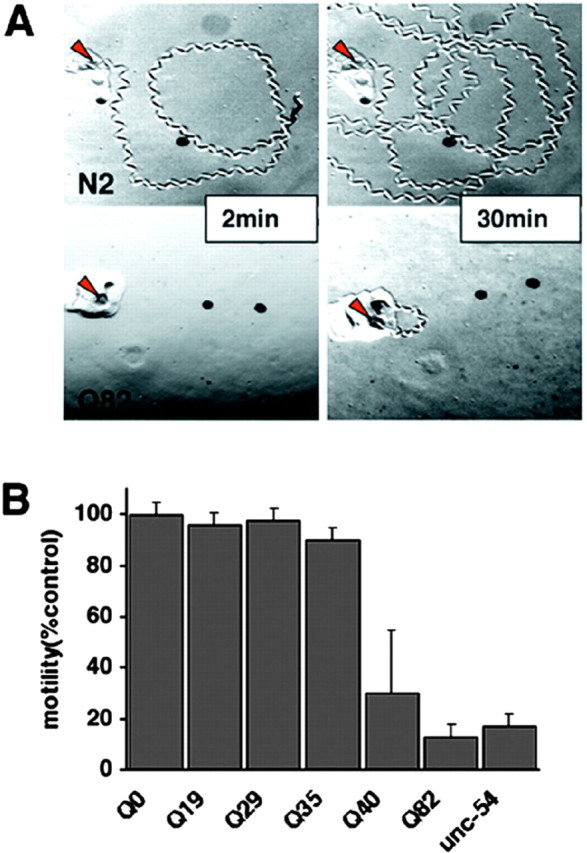
Expression of polyQ expansions in C. elegans muscle results in a motility defect that directly corresponds to aggregate formation. A, Time-lapse micrographs illustrating tracks left by 5-d-old wild-type (N2) and Q82 animals 2 and 30 min after being placed at the position marked by the red arrow. B, Quantitation of motility index for 4- to 5-d-old Q0, Q19, Q29, Q35, Q40, Q82, and unc-54(r293) animals. Data are mean ± SD for at least 50 animals of each type as a percentage of N2 motility. [Adapted from Morley et al. (2002).].
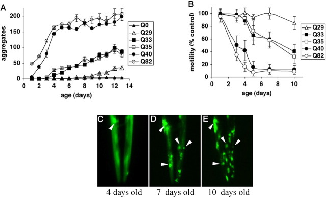
Influence of aging on polyQ aggregation and toxicity. A, Accumulation of aggregates in Q82 (○), Q40 (●), Q35 (□), Q33 (■), Q29 (▵), and Q0 (▴) during aging. Data are mean ± SEM. Twenty-four animals of each type are represented at day 1. Cohort sizes decreased as animals died during the experiment, but each data point represents at least five animals. B, Motility index as a function of age for the same cohorts of animals described in A. Data are mean ± SD as a percentage of age-matched Q0 animals. C–E, Epifluorescence micrographs of the head of an individual Q35 animal at 4 (C), 7 (D), and 10 (E) days of age, illustrating age-dependent accumulation of aggregates. Arrowheads indicate positions of the same aggregates on different days. In E, the animal is rotated slightly relative to its position in D. [Adapted from Morley et al. (2002).].
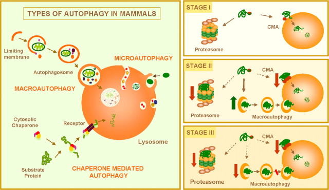
Autophagy and neurodegenerative disorders. Left, Mammalian cells use three types of autophagy. Microautophagy and macroautophagy involve the sequestration of complete cytosolic regions directly by the lysosomes or in an intermediate compartment, the autophagic vacuole, which is then delivered to lysosomes. In chaperone-mediated autophagy, single soluble proteins are recognized by a cytosolic chaperone and a receptor at the lysosomal membrane that mediates their translocation across the membrane into the lysosomal lumen. Right, Misfolded or altered proteins are selectively degraded by the ubiquitin/proteasome system or by chaperone-mediated autophagy (STAGE I). However, when these altered proteins organize in toxic multimeric complexes, they often alter the proteolytic activity of these two pathways. Upregulation of macroautophagy can compensate for this deficit (STAGE II). Aggravating factors, such as oxidative stress and aging, can precipitate the failure of macroautophagy with the consequent detrimental effect on cell functioning, often resulting in cellular death (STAGE III).
References
-
- Arrasate M, Mitra S, Schweitzer ES, Segal MR, Finkbeiner S. Inclusion body formation reduces levels of mutant huntingtin and the risk of neuronal death. Nature. 2004;431:805–810. - PubMed
-
- Beal MF, Ferrante RJ. Experimental therapeutics in transgenic mouse models of Huntington's disease. Nat Rev Neurosci. 2004;5:373–384. - PubMed
-
- Beal MF, Kowall NW, Ellison DW, Mazurek MF, Swartz KJ, Martin JB. Replication of the neurochemical characteristics of Huntington's disease by quinolinic acid. Nature. 1986;321:168–171. - PubMed
Publication types
MeSH terms
LinkOut - more resources
Full Text Sources
Other Literature Sources
Medical