

Review
doi: 10.1523/JNEUROSCI.3194-06.2006.
Cellular excitability and the regulation of functional neuronal identity: from gene expression to neuromodulation
Affiliations
- PMID: 17035518
- PMCID: PMC6674680
- DOI: 10.1523/JNEUROSCI.3194-06.2006
Item in Clipboard
Review
Cellular excitability and the regulation of functional neuronal identity: from gene expression to neuromodulation
J Neurosci.
.
No abstract available
Figures
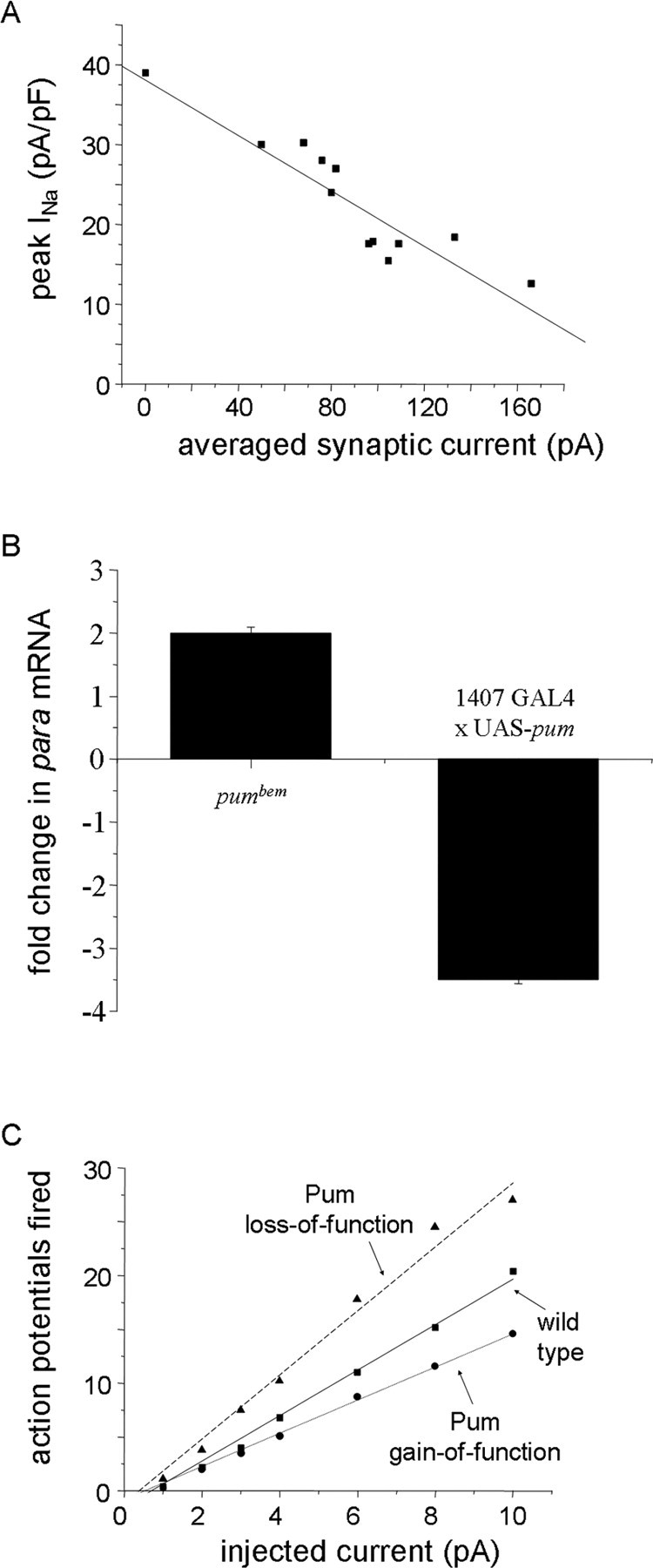
Exposure to synaptic excitation regulates INa and membrane excitability in Drosophila motoneurons. A, The amplitude of the voltage-gated INa recorded in Drosophila motoneurons is inversely related to the synaptic excitation that these neurons receive. B, The abundance of para mRNA in isolated CNS is significantly greater in the loss-of-function pumbem allele. By comparison, overexpression of UAS (upstream activating sequence)–pum in all of the neurons of the CNS (1407 GAL4) is sufficient to significantly reduce para mRNA. C, Membrane excitability, measured as action potential firing, shows a significant increase in Pumiliobem (loss-of-function) and a significant decrease after overexpression of UAS-pumilio (gain-of-function), respectively. The action potential firing rate was determined by injection of constant current (1–10 pA/500 ms) from a holding potential of −60 mV. A is reprinted from Baines and Pym (2006). B and C are reprinted from Mee et al. (2004).
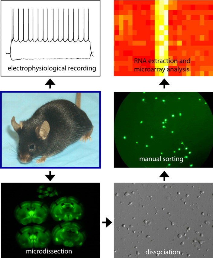
Mice in which subpopulations of neurons are genetically labeled are used for electrophysiological recordings and cell type-specific microarray analysis to obtain a correlated profile of firing properties and gene expression.
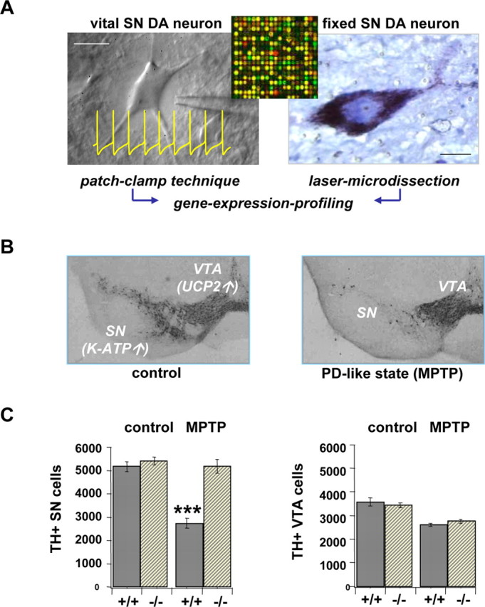
K-ATP channel activity triggers selective degeneration of SN DA neurons. A, A schematic overview of analyzing electrophysiological function and gene expression of individual dopaminergic neurons (mouse/human) combining brain slice patch clamping (inset, typical spontaneous activity of DA neurons) or laser microdissection with gene expression profiling (reverse transcription-PCR/microarrays). Scale bars, 15 μm. B, Dopaminergic neurons in SN and VTA [ marked by tyrosine hydroxylase (TH) immunostain] display differential vulnerability in the chronic mouse MPTP-model of PD. Differential mRNA levels of K-ATP channel subunits and UCP2 are indicated. C, Results of quantitative stereology indicated complete rescue of highly vulnerable SN DA neurons in the chronic MPTP PD model in a K-ATP channel knock-out mouse (Kir6.2 −/−; left). In contrast, MPTP-induced moderate loss of VTA DA neurons was independent from the presence (+ /+) or absence (−/−) of K-ATP channels (right).
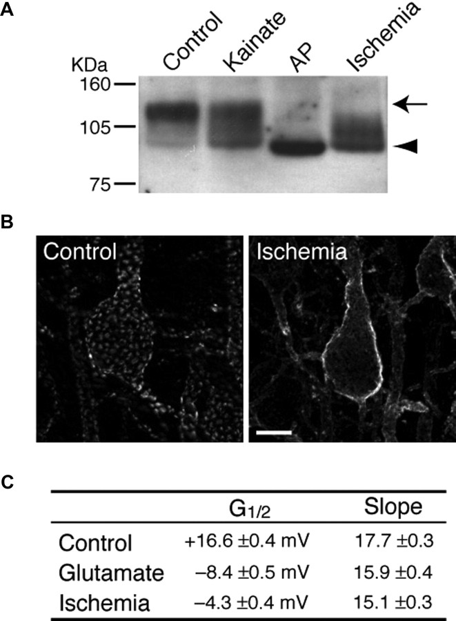
A, Dephosphorylation of Kv2.1 induced by seizures and ischemia. Rats were subjected to either kainate-induced continuous seizures (Kainate) or CO2-induced brain ischemia (Ischemia). Total brain membrane was prepared, and a fraction was treated in vitro with alkaline phosphatase (AP) to completely dephosphorylate Kv2.1. The arrow indicates the highly phosphorylated Kv2.1, and arrowhead indicates fully dephosphorylated Kv2.1. Numbers on the left denote the molecular weight standards. B, Dispersion of Kv2.1 clusters in brain by ischemia. Brain sections from control and ischemic rats were stained with a specific anti-Kv2.1 antibody. Images show pyramidal neurons in the cerebral cortex. It should be noted that kainate-induced seizures also resulted in dispersion of Kv2.1 clusters. Scale bar, 10 μm. C, Changes in the biophysical properties of IK/Kv2.1 by glutamate stimulation and ischemia in cultured neurons. IK currents were recorded in a cultured hippocampal neuron under whole-cell voltage clamp. The half-maximal activation membrane potential (G1/2) and the slope of the activation curve are shown (means ± SEM). The values are from Misonou et al. (2004, .
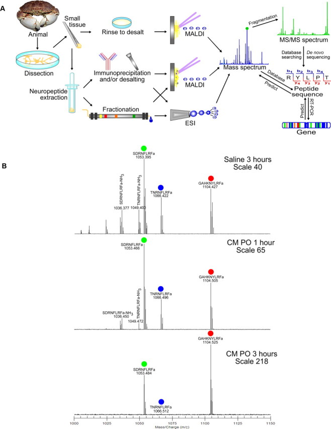
Development of a mass spectrometry-based peptidomic approach for understanding functional consequence of neuropeptide multiplicity. A, An overview of mass spectrometry-based strategy for neuropeptide discovery and characterization. B, Differential degradation of three FLRFamide (Phe-Leu-Arg-Phe-NH2) peptides by peptidase located in the pericardial organ monitored by matrix-assisted laser desorption/ionization Fourier transform mass spectrometry. A is reprinted from DeKeyser and Li (2006). B is adapted from Cruz-Bermudez et al. (2006). MS, Mass spectrometry; MALDI, matrix-assisted laser desorption/ionization; ESI, electrospray ionization; RT, reverse transcription; CM PO, Cancer magister pericardial organ.
Similar articles
-
Neurotransmitter regulation of neuronal outgrowth, plasticity and survival in the year 2001.Trends Neurosci. 1995 Feb;18(2):71-2. doi: 10.1016/0166-2236(95)93877-z. Trends Neurosci. 1995. PMID: 7537415 Review. No abstract available.
-
Development of sympathetic neurons: neurotransmitter plasticity and differentiation factors.Prog Brain Res. 1994;100:19-23. doi: 10.1016/s0079-6123(08)60763-3. Prog Brain Res. 1994. PMID: 7938519 Review. No abstract available.
-
Neurotransmitter Switching? No Surprise.Neuron. 2015 Jun 3;86(5):1131-44. doi: 10.1016/j.neuron.2015.05.028. Neuron. 2015. PMID: 26050033 Free PMC article. Review.
-
Reserve pool neuron transmitter respecification: Novel neuroplasticity.Dev Neurobiol. 2012 Apr;72(4):465-74. doi: 10.1002/dneu.20920. Dev Neurobiol. 2012. PMID: 21595049 Free PMC article. Review.
-
Neuromodulation as a mechanism for the induction of repetition priming.Curr Opin Neurobiol. 2014 Dec;29:33-8. doi: 10.1016/j.conb.2014.04.011. Epub 2014 May 16. Curr Opin Neurobiol. 2014. PMID: 25261622 Free PMC article. Review.
Cited by
-
Reduced Neural Excitability and Activation Contribute to Clinically Meaningful Weakness in Older Adults.J Gerontol A Biol Sci Med Sci. 2021 Mar 31;76(4):692-702. doi: 10.1093/gerona/glaa157. J Gerontol A Biol Sci Med Sci. 2021. PMID: 32588058 Free PMC article.
-
Canard solutions in neural mass models: consequences on critical regimes.J Math Neurosci. 2021 Sep 16;11(1):11. doi: 10.1186/s13408-021-00109-z. J Math Neurosci. 2021. PMID: 34529192 Free PMC article.
-
Rapid homeostatic plasticity of intrinsic excitability in a central pattern generator network stabilizes functional neural network output.J Neurosci. 2012 Jul 11;32(28):9649-58. doi: 10.1523/JNEUROSCI.1945-12.2012. J Neurosci. 2012. PMID: 22787050 Free PMC article.
-
Blurring the boundaries: developmental and activity-dependent determinants of neural circuits.Trends Neurosci. 2013 Oct;36(10):610-9. doi: 10.1016/j.tins.2013.06.006. Epub 2013 Jul 19. Trends Neurosci. 2013. PMID: 23876426 Free PMC article. Review.
-
Intracellular gold nanoparticles increase neuronal excitability and aggravate seizure activity in the mouse brain.PLoS One. 2014 Mar 13;9(3):e91360. doi: 10.1371/journal.pone.0091360. eCollection 2014. PLoS One. 2014. PMID: 24625829 Free PMC article.
References
-
- Andrews ZB, Diano S, Horvath TL. Mitochondrial uncoupling proteins in the CNS: in support of function and survival. Nat Rev Neurosci. 2005;6:829–840. - PubMed
-
- Baines RA. Neuronal homeostasis through translational control. Mol Neurobiol. 2005;32:113–121. - PubMed
-
- Baines RA, Pym EC. Determinants of electrical properties in developing neurons. Semin Cell Dev Biol. 2006;17:12–19. - PubMed
-
- Brezina V, Weiss KR. Functional consequences of divergence and convergence in physiological signaling pathways. Mol Psychiatry. 1997;2:9–11. - PubMed
Publication types
MeSH terms
Substances
LinkOut - more resources
Full Text Sources
Molecular Biology Databases