Comparative Study
. 2006 Oct 11;26(41):10397-406.
doi: 10.1523/JNEUROSCI.1671-06.2006.

Functional analyses of glycyl-tRNA synthetase mutations suggest a key role for tRNA-charging enzymes in peripheral axons

Affiliations
Comparative Study

Functional analyses of glycyl-tRNA synthetase mutations suggest a key role for tRNA-charging enzymes in peripheral axons

Anthony Antonellis et al. J Neurosci. .

Abstract

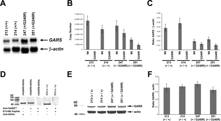
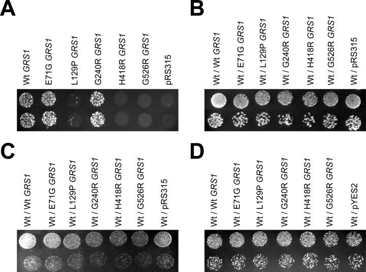
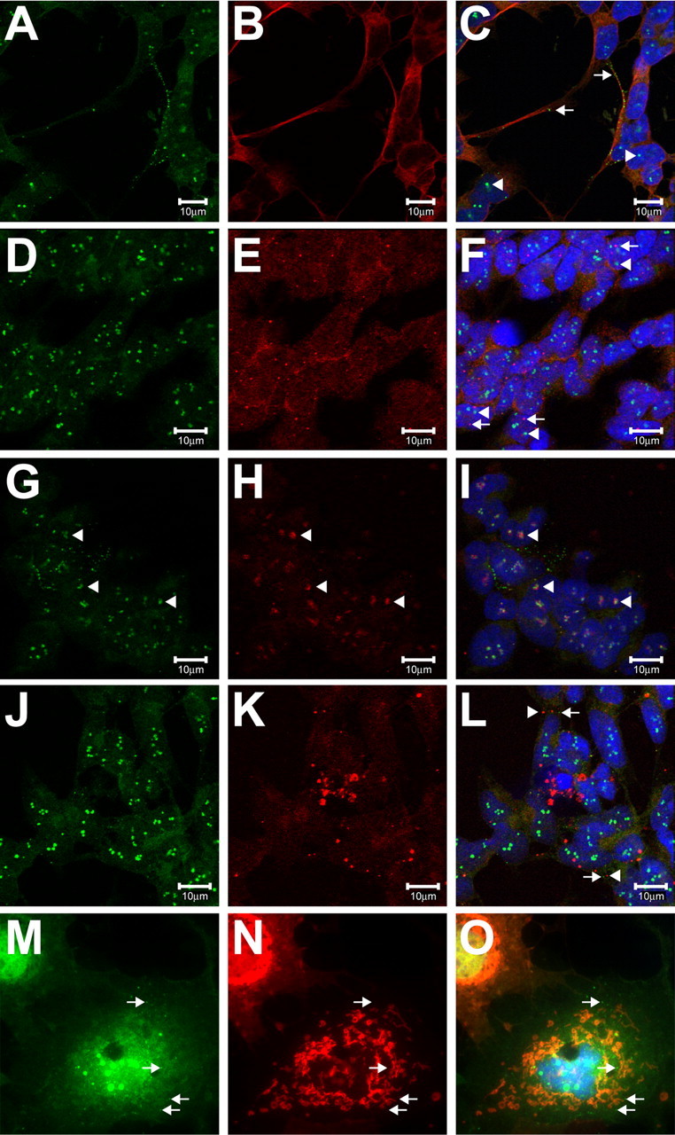
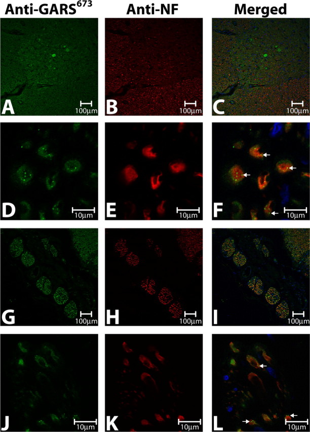
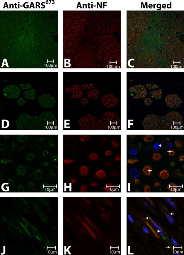
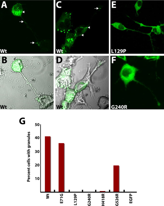
Charcot-Marie-Tooth disease type 2D (CMT2D) and distal spinal muscular atrophy type V (dSMA-V) are axonal neuropathies characterized by a phenotype that is more severe in the upper extremities. We previously implicated mutations in the gene encoding glycyl-tRNA synthetase (GARS) as the cause of CMT2D and dSMA-V. GARS is a member of the family of aminoacyl-tRNA synthetases responsible for charging tRNA with cognate amino acids; GARS ligates glycine to tRNA(Gly). Here, we present functional analyses of disease-associated GARS mutations and show that there are not any significant mutation-associated changes in GARS expression levels; that the majority of identified GARS mutations modeled in yeast severely impair viability; and that, in most cases, mutant GARS protein mislocalizes in neuronal cells. Indeed, four of the five mutations studied show loss-of-function features in at least one assay, suggesting that tRNA-charging deficits play a role in disease pathogenesis. Finally, we detected endogenous GARS-associated granules in the neurite projections of cultured neurons and in the peripheral nerve axons of normal human tissue. These data are particularly important in light of the recent identification of CMT-associated mutations in another tRNA synthetase gene [YARS (tyrosyl-tRNA synthetase gene)]. Together, these findings suggest that tRNA-charging enzymes play a key role in maintaining peripheral axons.


Figures

Figure 1.
Figure 1.
Expression of mutant GARS. A, Total RNA was extracted from lymphoblastoid cell lines derived from unaffected noncarriers (+/+) and CMT2D-affected carriers of the G240R GARS mutation (+/G240R) and subjected to Northern blot analysis. Blots were hybridized with a GARS- or β-actin-specific probe, as indicated. B, Real-time PCR was performed using reverse-transcribed RNA from the cell lines described in A and allele-specific primers. Copy number of the wild-type (Wt) and G240R alleles was measured by including known concentrations of control plasmids containing wild-type or G240R GARS mRNA. C, Real-time PCR studies were performed similarly to those in B, except that primers specific for the β-actin gene were included to assess the efficiency of the reverse-transcription reactions. The ratio of GARS mRNA to β-actin mRNA was then calculated. D, rhGARS–6XHis was subjected to Western blot analysis using an anti-GARS673 or anti-6XHis antibody, as indicated. In two cases, the hybridization was performed in the presence of excess 673–685 peptide. Similar analyses were performed with a total protein lysate from a lymphoblastoid cell line of an unaffected noncarrier (+/+) using anti-GARS673. E, Western blot analyses were performed with total protein lysates of lymphoblastoid cell lines from unaffected noncarriers (+/+) and CMT2D-affected carriers of the G240R GARS mutation (+/G240R) using an anti-GARS673 or anti-actin antibody, as indicated. F, The ratio of GARS to actin protein levels was determined by dividing the optical density of each GARS-specific Western blot band with that of the corresponding actin-specific band (e.g., as in E) using four independent Western blots. Error bars indicate SEM.
Figure 2.
Figure 2.
Growth of yeast strains containing wild-type and mutant GRS1. A, Representative cultures of the indicated yeast strains were inoculated and grown on medium containing 5-FOA. Each strain had been transfected previously with a vector containing no insert (pRS315), wild-type (Wt) GRS1, or the indicated mutant form of GRS1 that modeled a human GARS mutation (Table 1). B, Representative cultures of the indicated yeast strains were inoculated and grown on medium containing glucose. Each strain contains the wild-type GRS1-bearing maintenance vector (see Materials and Methods for details) as well as a transformed vector expressing the indicated GRS1 allele from the endogenous promoter. The Wt/pRS315 strain contained a transformed vector without an insert. C, Similar experiment to that in B, except that strains were grown on medium containing glycerol. D, Representative cultures of the indicated strains were inoculated and grown on medium containing galactose; each strain contained two copies of GRS1, one wild-type copy expressed from the endogenous promoter and one expressing the indicated allele from the GAL1 promoter.
Figure 3.
Figure 3.
Expression of endogenous GARS in cultured cells. A, B, D, E, G, H, J, K, Differentiated human SH-SY5Y cells were stained with anti-GARS673 (A, D, G, J), anti-α-tubulin (B), anti-SMN (E), anti-nucleolar antigen (H), anti-OxPhos complex IV (K), and DAPI and examined by confocal fluorescence microscopy. C, F, I, L, Merged images depict large GARS-associated globules in the nuclei (C, arrowheads) and granules in the neurite projections (C, arrows); distinct and separate localization of SMN gems (F, arrows) and nuclear GARS-associated globules (F, arrowheads); multiple sites of colocalized GARS-associated globules and nucleoli in the nucleus (I, arrowheads); and distinct cytoplasmic GARS-associated granules (L, arrows) in the absence of mitochondrial staining (L, arrowheads). M–O, COS7 cells were stained with anti-GARS673 and DAPI in the absence (M) or presence (N) of the transfected pDsRed2–Mito vector. The merged image (O) shows GARS-associated granules (arrows) distinct from the labeled mitochondria.
Figure 4.
Figure 4.
Localization of GARS in the ventral horn and ventral root. A, B, D, E, G, H, J, K, Human thoracic spinal-cord sections were stained with anti-GARS673 (A, D, G, J), anti-neurofilament (anti-NF; B, E, H, K), and DAPI and examined by confocal fluorescence microscopy. C, F, I, L, Merged images show strong GARS staining in the ventral horn (C), GARS-associated granules in ventral horn axons (F, arrows), strong GARS staining in ventral root axons (I), and GARS-associated granules in ventral root axons (L, arrows).
Figure 5.
Figure 5.
Localization of GARS in the dorsal horn, dorsal root, and sural nerve. Human thoracic spinal cord (A–I) and sural nerve (J–L) sections were stained with anti-GARS673 (A, D, G, J), anti-neurofilament (anti-NF; B, E, H, K), and DAPI and examined by confocal fluorescence microscopy. Merged images show strong GARS staining in the dorsal horn (C), strong GARS staining in dorsal root axons (F), and GARS-associated granules in dorsal root (I, arrows) and sural nerve (L, arrows) axons. Note that GARS staining can also be seen in nuclei of the dorsal root and sural nerve (I, L, arrowheads).
Figure 6.
Figure 6.
Expression of wild-type and mutant GARS–EGFP in mouse motor neuron cells. A, C, E, F, Differentiated MN-1 cells expressing wild-type (Wt) GARS–EGFP (A, C), L129P GARS–EGFP (E), or G240R GARS–EGFP (F) were examined by fluorescence microscopy. Wild-type GARS–EGFP-associated granules within the cell body and neurite projections are indicated by arrowheads and arrows, respectively. B, D, Merged images from fluorescence and differential interference contrast microscopy reveal cell morphology and neurite-projection paths of cells expressing wild-type GARS–EGFP. G, The percentage of EGFP-positive cells containing GARS-associated granules was determined after transfection with constructs expressing wild-type GARS–EGFP, the indicated mutant forms of GARS–EGFP, or EGFP alone. Five replicate experiments consistently gave the observed results.

References

    1. Antonellis A, Ellsworth RE, Sambuughin N, Puls I, Abel A, Lee-Lin SQ, Jordanova A, Kremensky I, Christodoulou K, Middleton LT, Sivakumar K, Ionasescu V, Funalot B, Vance JM, Goldfarb LG, Fischbeck KH, Green ED. Glycyl tRNA synthetase mutations in Charcot–Marie–Tooth disease type 2D and distal spinal muscular atrophy type V. Am J Hum Genet. 2003;72:1293–1299. - PMC - PubMed
    1. Biedler JL, Roffler-Tarlov S, Schachner M, Freedman LS. Multiple neurotransmitter synthesis by human neuroblastoma cell lines and clones. Cancer Res. 1978;38:3751–3757. - PubMed
    1. Boeke JD, LaCroute F, Fink GR. A positive selection for mutants lacking orotidine-5′-phosphate decarboxylase activity in yeast: 5-fluoro-orotic acid resistance. Mol Gen Genet. 1984;197:345–346. - PubMed
    1. Christodoulou K, Kyriakides T, Hristova AH, Georgiou DM, Kalaydjieva L, Yshpekova B, Ivanova T, Weber JL, Middleton LT. Mapping of a distal form of spinal muscular atrophy with upper limb predominance to chromosome 7p. Hum Mol Genet. 1995;4:1629–1632. - PubMed
    1. Corti O, Hampe C, Koutnikova H, Darios F, Jacquier S, Prigent A, Robinson JC, Pradier L, Ruberg M, Mirande M, Hirsch E, Rooney T, Fournier A, Brice A. The p38 subunit of the aminoacyl-tRNA synthetase complex is a Parkin substrate: linking protein biosynthesis and neurodegeneration. Hum Mol Genet. 2003;12:1427–1437. - PubMed

Publication types

Substances