. 2006 Oct 18;25(20):4843-53.
doi: 10.1038/sj.emboj.7601364. Epub 2006 Oct 12.

Epigenetic determination of a cell-specific gene expression program by ATF-2 and the histone variant macroH2A

Affiliations

Epigenetic determination of a cell-specific gene expression program by ATF-2 and the histone variant macroH2A

Marios Agelopoulos et al. EMBO J. .

Abstract

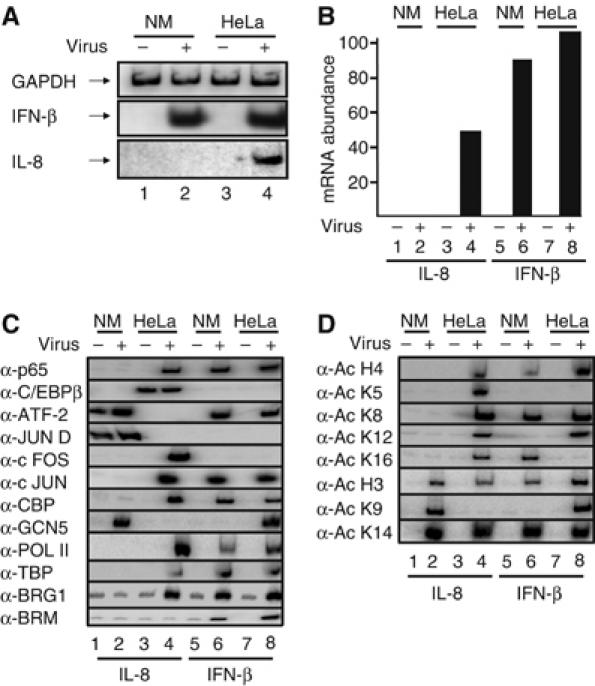
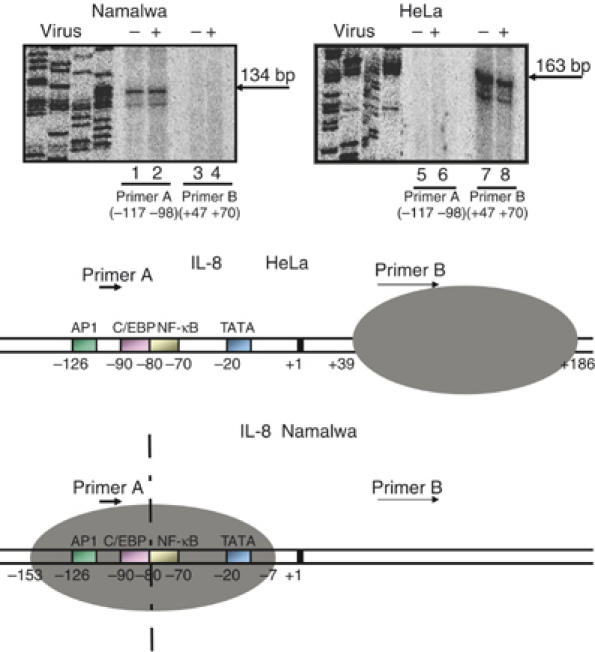
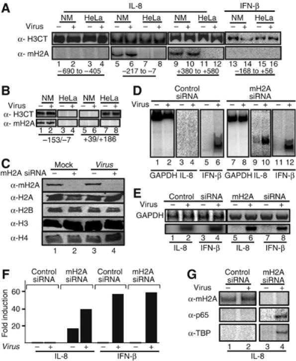
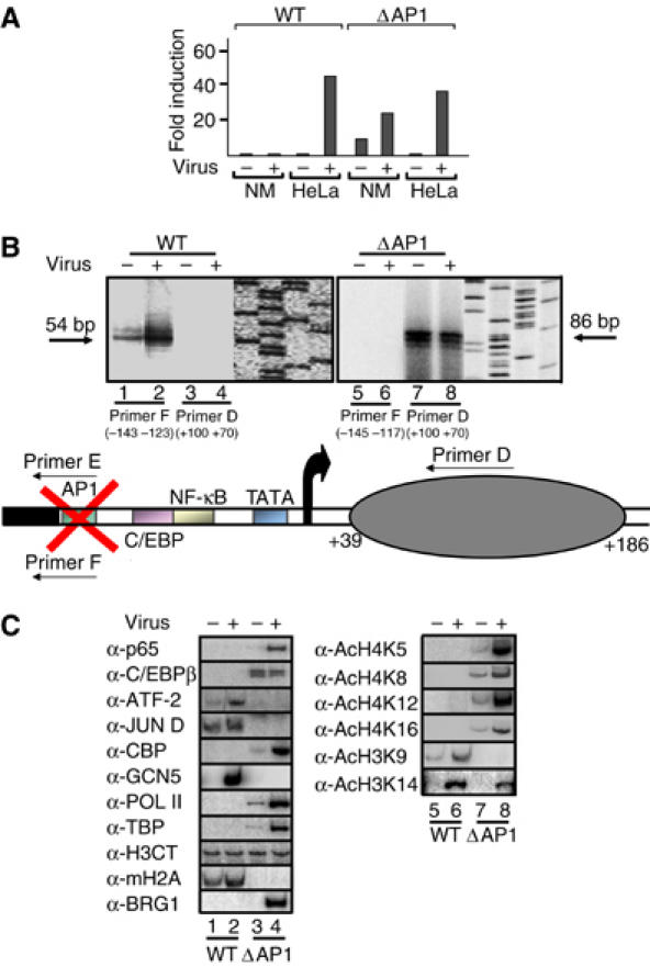
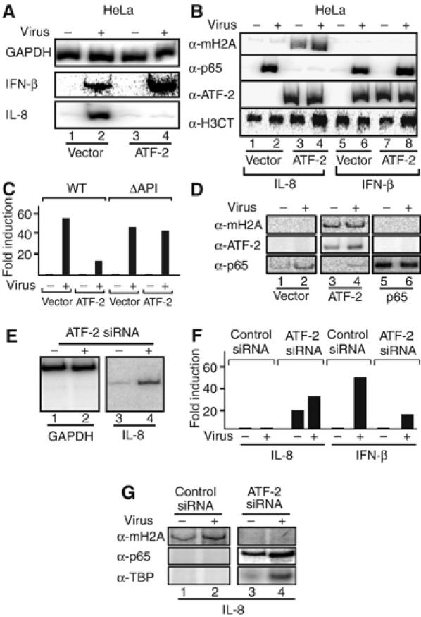
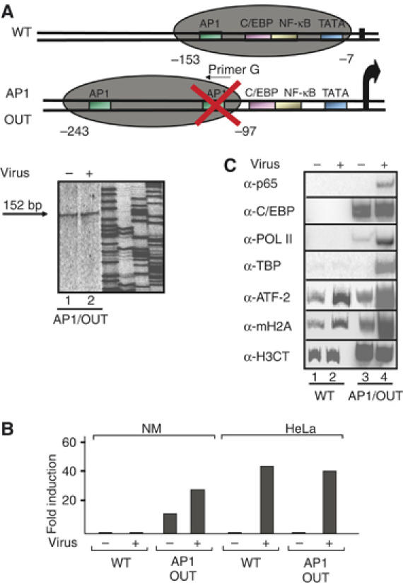
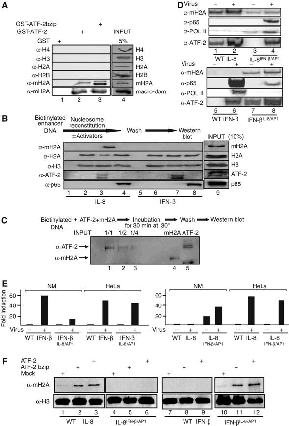
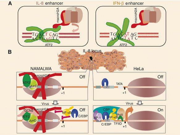
Transcriptional activation of the interleukin-8 (IL-8) gene is restricted to distinct cell types, although the transcriptional regulatory proteins controlling IL-8 gene expression are ubiquitous. We show that cell-specific transcription of IL-8 is due to the distinct chromatin architecture on the enhancer/promoter before the arrival of the inducing signal. In expressing epithelial cells the enhancer/promoter is nucleosome-free, whereas in non-expressing B cells a nucleosome masks the entire regulatory region. The B-cell-specific nucleosome contains the histone variant macroH2A, which is responsible for preventing transcription factor binding. Recruitment of the repressive macroH2A nucleosome requires direct interactions between ATF-2 bound to the nearby AP1 site and macroH2A and it is regulated by DNA-induced protein allostery. siRNA against ATF-2 or macroH2A rescues IL-8 transcription in B cells. Thus, a transcription factor can work as a transcriptional repressor by orchestrating and maintaining the assembly of specialized local chromatin architectures.


Figures

Figure 1
Figure 1
Cell-type-specific recruitment of transcriptional regulatory proteins to the IL-8 enhancer/promoter. (A) Total RNA was isolated from mock- or virus-infected HeLa and Namalwa (NM) cells for 6 h, and used in RT–PCR reactions as a template with primers detecting the transcripts shown on the left part of the gel. (B) Real-time RT–PCR analysis illustrating cell-type-specific transcriptional activation of the IL-8 gene. (C) Crosslinked chromatin prepared from mock or 6 h virus-infected HeLa and NM cells was immunoprecipitated with the indicated antibodies against transcriptional regulatory proteins required for IL-8 or IFN-β gene expression. The precipitated IL-8 and IFN-β promoters were detected by PCR using 32P-dCTP in the reaction. (D) Same as in (C) except the antibodies used reacted with the indicated acetylated histone lysine residues.
Figure 2
Figure 2
The chromatin architecture of the IL-8 gene enhancer/promoter differs between distinct cell types. A diagrammatic illustration depicting the distinct chromatin architecture of the IL-8 enhancer/promoter in Namalwa and HeLa cells is shown at the bottom of the figure. The dotted vertical line indicates the axis of dyad symmetry of the nucleosome in Namalwa cells. HeLa and Namalwa cells were mock or virus infected for 6 h followed by formaldehyde crosslinking and micrococcal nuclease treatment of the isolated chromatin. The DNA was purified and annealed with primers A and B followed by extension and ligation-mediated PCR using 32P-dCTP in the reaction to detect the extended products. A sequencing gel containing the extended products run side-by-side with the DNA sequencing reactions serving as size markers is shown.
Figure 3
Figure 3
The histone variant macroH2A marks the IL-8 gene for repression in B cells. (A) HeLa and Namalwa cells were mock or virus infected for 6 h followed by formaldehyde crosslinking and immunoprecipitation using antibodies specific for macroH2A1.2 and the carboxyl-terminus of histone H3. The precipitated chromatin-bound DNA was detected using the indicated set of primers amplifying the upstream IL-8 region (−690 to −405, lanes 1–4), the enhancer/promoter (−217 to −7, lanes 5–8) and the coding region (+380 to +580, lanes 9–12) or the IFN-β enhancer (lanes 13–16). The relative position of the amplified regions is shown at the bottom of the gel. (B) Same as in (A) except the chromatin used was composed of mononucleosomes prepared after digestion with micrococcal nuclease. (C) A Western blot using whole-cell extracts prepared from mock- or virus-infected HeLa cells transfected with a control siRNA (lanes 1, 3) or the macroH2A-specific siRNA (lanes 2 and 4). Our transfection efficiency in HeLa cells is approximately 90%. The abundance of macroH2A1.2 and histones H2A, H2B, H3 and H4 was determined by Western blotting using specific antibodies. (D) Namalwa cells were transfected with the control (lanes 1–6) or the macroH2A-specific siRNA (lanes 7–12) and 48 h later they were infected with Sendai virus for 6 h. The isolated RNA was used as a template in RT–PCR reactions using primers detecting GAPDH, IL-8 and the IFN-β-coding regions. (E) Same as in (D) except the RNAs used in the RT–PCR reactions were prepared from HeLa cells transfected with the indicated siRNAs. (F) Namalwa cells were transfected with luciferase-based reporter plasmids bearing the IL-8 or the IFN-β enhancer promoter along with control or macroH2A siRNAs. At 48 h following transfection, the cells were mock or virus infected for 10 h and the luciferase activity was determined. The average of three experiments is shown and the variability from experiment to experiment was less than 20%. (G) Namalwa cells were transfected with the IL-8 reporter as in (F). At 6 h after virus infection, the cells crosslinked with formaldehyde and the isolated chromatin was immunoprecipitated using the indicated antibodies. PCR was used to detect the precipitated IL-8 promoter fragments using plasmid-specific primers.
Figure 4
Figure 4
The ATF/AP1 site of the IL-8 enhancer is required for the recruitment and positioning of the macroH2A-containing repressing nucleosome. (A) HeLa and Namalwa cells were transfected with either the WT or the ΔAP1 IL-8 enhancer/promoter-luciferase fusion constructs followed by virus infection for 10 h. Shown is the average of seven experiments and the variation from experiment to experiment was less than 35%. (B) Namalwa cells were transfected with the WT (−127/+59) (lanes 1–4) or an isogenic IL-8 enhancer/promoter construct bearing a mutation in the ATF/AP1 site (ΔAP1) (lanes 5–8) followed by mock or virus infection. Mononucleosomal DNA was purified and annealed with the indicated primers followed by ligation-mediated PCR. To distinguish between the endogenous and the exogenous WT and mutant templates, we used promoter-luciferase fusion constructs. Thus, primer D hybridizes to luciferase sequences and therefore detects the exogenous genes only. Primer E hybridizes to the mutant AP1 site only and the nearby vector sequences, whereas primer F recognizes the WT AP1 site and the nearby vector sequences. The diagrammatic illustration at the bottom of the figure illustrates the position of the nucleosome in the ΔAP1 template in Namalwa cells. (C) The WT and the ΔAP1 IL-8 enhancer/promoter templates were transfected into Namalwa cells followed by virus infection for 6 h. Crosslinked chromatin was immunoprecipitated with the indicated antibodies and the IL-8 enhancer/promoter was detected by PCR using vector-specific primers and 32P-dCTP in the reaction.
Figure 5
Figure 5
ATF-2 functions as a repressor of IL-8 transcription. (A) HeLa cells were transfected with empty or ATF-2-expressing vector and 40 h following transfection the cells were infected with Sendai virus for 6 h. RNA was prepared and the abundance of GAPDH, IL-8 and IFN-β mRNAs was determined by RT–PCR. (B) HeLa cells were transfected and infected with Sendai virus as in (A). Following virus infection, the cells were crosslinked with formaldehyde and the isolated chromatin was precipitated with the indicated antibodies. PCR was used to detect the precipitated endogenous IL-8 and IFN-β promoters. (C) HeLa cells were transfected with the WT or the ΔAP1 IL-8 enhancer/promoter luciferase fusion constructs and were mock or virus infected for 10 h. Shown is the average of three experiments and the variability from experiment to experiment was less than 25%. (D) HeLa cells were transfected with the WT IL-8 luciferase reporter along with empty (lanes 1 and 2) or ATF-2 (lanes 3 and 4) or p65-expressing vectors (lanes 5 and 6). Following transfection, the cells were virus infected for 6 h, crosslinked with formaldehyde and the isolated chromatin was immunoprecipitated using the indicated antibodies. PCR was used to detect the precipitated IL-8 promoter fragments using plasmid-specific primers. (E) Namalwa cells were transfected with an siRNA against ATF-2 and 48 h later the cells were infected with Sendai virus followed by RNA isolation and RT–PCR analysis using IL-8-specific primers. (F) Namalwa cells were transfected with the IL-8 or IFN-β luciferase reporters along with control or ATF-2 siRNA. At 48 h following transfection, the cells were mock or virus infected for 8 h and the luciferase activity was determined. Shown is the average of two experiments and the variability from experiment to experiment was less than 25%. (G) Namalwa cells were transfected with the IL-8 reporter as in (F). At 6 h after virus infection, the cells were crosslinked with formaldehyde and the isolated chromatin was immunoprecipitated using the indicated antibodies. PCR was used to detect the precipitated IL-8 promoter fragments using plasmid-specific primers.
Figure 6
Figure 6
Altering the chromatin architecture of the IL-8 promoter by relocating the ATF site. (A) Namalwa cells were transfected with the AP1 OUT IL-8 reporter followed by isolation of mononucleosomes. The DNA was purified and annealed with the indicated primer G (hybridizes to the mutated AP1 site) followed by ligation-mediated PCR. The diagrammatic illustration at the top of the figure illustrates the position of the nucleosome in the AP1 OUT as opposed to the WT template in Namalwa cells. (B) Namalwa cells were transfected with the WT or the AP1 OUT IL-8 templates and 48 h later the cells were infected with Sendai virus for 10 h followed by determination of the luciferase activity. Shown is the average of four experiments and the variability from experiment to experiment was less than 30%. (C) Namalwa cells were transfected with the IL-8 reporters as in (B). At 6 h after virus infection, the cells were crosslinked with formaldehyde and the isolated chromatin was immunoprecipitated using the indicated antibodies. PCR was used to detect the precipitated IL-8 promoter fragments using plasmid-specific primers.
Figure 7
Figure 7
Direct recruitment of macroH2A nucleosomes to the IL-8 enhancer by ATF-2. (A) Glutathione beads containing immobilized GST, GST-ATF2 or GST-ATF2bzip were incubated with recombinant and purified histones H4, H3, H2A, H2B, macroH2A or the macro-domain of the macroH2A. Bound proteins were detected by Western blots using the indicated antibodies. (B) Biotinylated IL-8 (lanes 1–4) or IFN-β (lanes 5–8) promoter fragments were attached to Dyna beads and reconstituted into chromatin using HeLa donor chromatin in the absence (lanes 2 and 6) or in the presence of recombinant ATF-2 (lanes 3 and 7) or p65 (lanes 4 and 8). Lanes 1 and 5 correspond to beads only. Next, the fragments were washed three times before Western blot analysis using the indicated antibodies. (C) A biotinylated IL-8 enhancer/promoter DNA fragment was incubated with the indicated amounts of ATF-2 and free recombinant macroH2A followed by washes and Western blot. The last two lanes on the right depict the amounts of proteins used as input in the experiment (1/1). (D) Namalwa cells were transfected as in (E). At 6 h after virus infection, the cells were crosslinked with formaldehyde and the isolated chromatin was immunoprecipitated using the indicated antibodies. PCR was used to detect the precipitated IL-8 and IFN-β promoter fragments using plasmid-specific primers. (E) Luciferase-based reporter constructs bearing the WT IL-8 and IFN-β and their swapped derivatives were transfected into HeLa or Namalwa cells followed by virus infection. The corresponding luciferase activities were determined from three independent experiments with less than 35% variability from experiment to experiment. (F) Biotinylated IL-8 (lanes 1–3), IL-8IFNβ/AP1 (lanes 4–6), IFN-β (lanes 7–9) or IFNβIL-8/AP1 (lanes 10–12) promoter fragments were attached to Dyna beads and reconstituted into chromatin using HeLa donor chromatin in the absence (lanes 1, 4, 7 and 10) or in the presence of recombinant ATF-2 bzip (lanes 2, 5, 8 and 11) or full-length ATF-2 (lanes 3, 6, 9 and 12). Next, the fragments were washed three times before Western blot analysis using the indicated antibodies.
Figure 8
Figure 8
A model for the cell-type-specific expression of the IL-8 gene. (A) A model depicting distinct configurations of ATF-2 bzip domain bound to the IL-8 and IFN-β sites, correlating with the recruitment of macoH2A-containing nucleosomes. (B) The default chromatin architecture of the IL-8 enhancer/promoter differs between distinct cell types. Namalwa B cells are incapable of inducing the gene in response to virus infection because a nucleosome containing the histone variant macroH2A masks the enhancer/promoter region and inhibits the association of the IL-8 activators. This nucleosome is recruited via direct protein–protein interactions between the constitutively bound ATF-2/JunD heterodimer and the macro-domain of macroH2A. In contrast, ATF-2/JunD don not associate with the IL-8 enhancer in HeLa cells and therefore the macroH2A nucleosome is not recruited, thus allowing transcriptional activation.

References

    1. Abbott DW, Chadwick BP, Thambirajah A, Ausio J (2005) Beyond the Xi: macroH2A chromatin distribution and post-translational modification in an avian system. J Biol Chem 280: 16437–16445 - PubMed
    1. Abbott DW, Laszczak M, Lewis JD, Su H, Moore SC, Hills M, Dimitrov S, Ausio J (2004) Structural characterization of macroH2A containing chromatin. Biochemistry 43: 1352–1359 - PubMed
    1. Agalioti T, Chen G, Thanos D (2002) Deciphering the transcriptional histone acetylation code for a human gene. Cell 111: 381–392 - PubMed
    1. Angelov D, Molla A, Perche PY, Hans F, Cote J, Khochbin S, Bouvet P, Dimitrov S (2003) The histone variant macroH2A interferes with transcription factor binding and SWI/SNF nucleosome remodeling. Mol Cell 11: 1033–1041 - PubMed
    1. Chakravarthy S, Gundimella SK, Caron C, Perche PY, Pehrson JR, Khochbin S, Luger K (2005) Structural characterization of the histone variant macroH2A. Mol Cell Biol 25: 7616–7624 - PMC - PubMed

Publication types

MeSH terms