Review
. 2006 Oct;29(10):1130-59.
doi: 10.1111/j.1540-8159.2006.00507.x.

Brugada syndrome

Affiliations
Review

Brugada syndrome

Charles Antzelevitch. Pacing Clin Electrophysiol. 2006 Oct.

Abstract

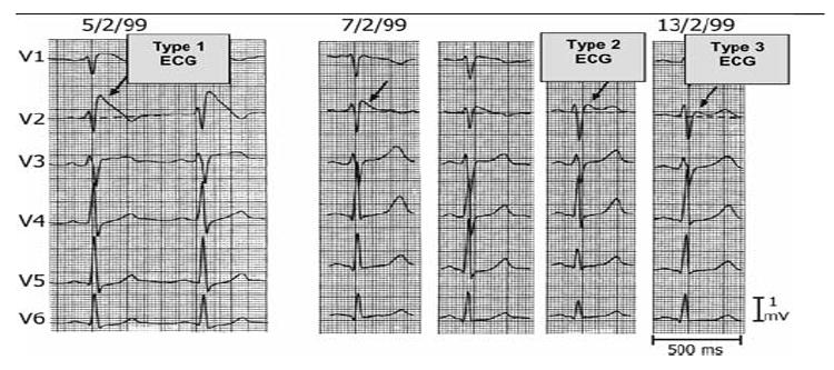
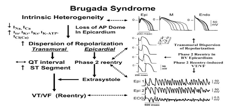
First introduced as a new clinical entity in 1992, the Brugada syndrome is associated with a relatively high risk of sudden death in young adults, and occasionally in children and infants. Recent years have witnessed a striking proliferation of papers dealing with the clinical and basic aspects of the disease. Characterized by a coved-type ST-segment elevation in the right precordial leads of the electrocardiogram (ECG), the Brugada syndrome has a genetic basis that thus far has been linked only to mutations in SCN5A, the gene that encodes the alpha-subunit of the sodium channel. The Brugada ECG is often concealed, but can be unmasked or modulated by a number of drugs and pathophysiological states including sodium channel blockers, a febrile state, vagotonic agents, tricyclic antidepressants, as well as cocaine and propranolol intoxication. Average age at the time of initial diagnosis or sudden death is 40 +/- 22, with the youngest patient diagnosed at 2 days of age and the oldest at 84 years. This review provides an overview of the clinical, genetic, molecular, and cellular aspects of the Brugada syndrome, incorporating the results of two recent consensus conferences. Controversies with regard to risk stratification and newly proposed pharmacologic strategies are discussed.


Figures

Figure 1
Figure 1
Three Types of ST-segment elevation generally observed in patients with the Brugada syndrome. Shown are precordial leads recorded from a patient diagnosed with the Brugada syndrome. Note the dynamic ECG changes occurring over a period of 2 days. The left panel shows a clear Type 1 ECG, which is diagnostic of the Brugada syndrome. A saddleback ST-segment elevation (Type 2) is observed on February 7,1999. The ST segment is further normalized on February 13, 1999, showing a Type 3 ECG. Modified from Wilde et al. with permission.
Figure 2
Figure 2
Shift of right precordial leads to 2nd and 3rd intercostal space unmasks a Type 1 Brugada ECG. (Top) Plot of 87 unipolar electrode sites (dots) and of 6 precordial electrocardiograms (ECG) (crosses). Eighty seven-lead points are arranged in a lattice-like pattern (13 × 7 matrix), except for four lead points on both mid-axillary lines and covered the entire thoracic surface. V1 and V2 leads of the ECG are located between D5 and E5, and between E5 and F5, respectively, whereas V4, V5, and V6 are coincident with G4, H4, and I4, respectively. (Bottom) Twelve-leads electrocardiograms (ECG) in a patient with Brugada syndrome. Type 2 saddleback ST-segment elevation was observed in V1 and V2 of the standard 12-lead ECG (4th intercostal space), whereas typical Type 1 coved-type ST-segment elevation was apparent in V1 and V2 recorded from the 2nd and 3rd intercostal space (arrows). Modified from Antzelevitch et al. , with permission.
Figure 3
Figure 3
Schematic of SCN5A, the gene that encodes the (-subunit of the sodium channel, illustrating mutations linked to Brugada syndrome, long-QT3 syndrome, conduction disease, and atrial standstill. Some mutations are associated with combined phenotypes. α = subunit
Figure 4
Figure 4
Phase 2 reentry. Reentrant activity induced by exposure of a canine ventricular epicardial preparation (0.7 cm2) to simulated ischemia. Microelectrode recordings were obtained from four sites as shown in the schematic (upper right). After 35 min of ischemia, the action potential dome develops normally at site 4, but not at sites 1, 2, or 3. The dome then propagates in a clockwise direction re-exciting sites 3, 2, and 1 with progressive delays, thus generating a closely coupled reentrant extrasystole (156 ms) at site 1. In this example of phase 2 reentry, propagation of the dome occurs in a direction opposite to that of phase 0, a mechanism akin to reflection. BCL = 700 ms. Modified from Lukas and Antzelevitch with permission.
Figure 5
Figure 5
ECG and arrhythmias with typical features of the Brugada syndrome recorded from canine right ventricular wedge preparations. (A) Schematic of arterially perfused right ventricular wedge preparation. (B) Pressure-induced phase 2 reentry and VT. Shown are transmembrane action potentials simultaneously recorded from two epicardial (Epi 1 and Epi 2) and one M region (M) sites, together with a transmural ECG. Local application of pressure near Epi 2 results in loss of the action potential dome at that site but not at Epi 1 or M sites. The dome at Epi 1 then re-excites Epi 2, giving rise to a phase 2 reentrant extrasystole which triggers a short run of ventricular tachycardia. Note the ST-segment elevation due to loss of the action potential dome in a segment of epicardium. (C) Polymorphic VT/VF induced by local application of the potassium channel opener pinacidil (10 μM) to the epicardial surface of the wedge. Action potentials from two epicardial sites (Epi 1 and Epi 2) and a transmural ECG were simultaneously recorded. Loss of the dome at Epi 1 but not Epi 2 creates a marked dispersion of repolarization, giving rise to a phase 2 reentrant extrasystole. The extrasystolic beat then triggers a long episode of ventricular fibrillation (22 sec). (Right panel) Addition of 4-aminopyridine (4-AP, 2 mM), a specific Ito blocker, to the perfusate restored the action potential dome at Epi 1, thus reducing dispersion of repolarization and suppressing all arrhythmic activity. BCL = 2,000 ms. (D) Phase 2 reentry gives rise to VT following addition of pinacidil (2.5 μM) to the coronary perfusate. Transmembrane action potentials form 2 epicardial sites (Epi 1 and Epi 2) and one endocardial site (Endo), as well as a transmural ECG were simultaneously recorded. (Right panel) 4-AP (1 mM) markedly reduces the magnitude of the action potential notch in epicardium, thus restoring the action potential dome throughout the preparation and abolishing all arrhythmic activity. Panel D is from Yan and Antzelevitch with permission.
Figure 6
Figure 6
Terfenadine-induced ST-segment elevation, T-wave inversion, transmural and endocardial dispersion of repolarization, and phase 2 reentry. Each panel shows transmembrane action potentials from one endocardial (top) and two epicardial sites together with a transmural ECG recorded from a canine arterially perfused right ventricular wedge preparation. (A) Control (BCL 400 ms). (B) Terfenadine (5 μM) accentuated the epicardial action potential notch creating a transmural voltage gradient that manifests as a ST-segment elevation or exaggerated J wave in the ECG. First beat recorded after changing from BCL 800 ms to BCL 400 ms. (C) Continued pacing at BCL 400 ms results in all-or-none repolarization at the end of phase 1 at some epicardial sites but not others, creating a local epicardial dispersion of repolarization (EDR) as well as a transmural dispersion of repolarization (TDR). (D) Phase 2 reentry occurs when the epicardial action potential dome propagates from a site where it is maintained to regions where it has been lost. Modified from Fish and Antzelevitch with permission.
Figure 7
Figure 7
Spontaneous and programmed electrical stimulationinduced polymorphic VT in RV wedge preparations pretreated with terfenadine (5–10 μM). (A) Phase 2 reentry in epicardium gives rise to a closely coupled extrasystole. (B) Phase 2 reentrant extrasystole triggers a brief episode of polymorphic VT. (C) Phase 2 reentry followed by a single circus movement reentry in epicardium gives rise to a couplet. (D) Extrastimulus (S1-S2 = 250 ms) applied to epicardium triggers a polymorphic VT. Modified from Fish and Antzelevitch with permission.
Figure 8
Figure 8
Schematic representation of right ventricular epicardial action potential changes proposed to underlie the electrocardiographic manifestation of the Brugada syndrome. Modified from Antzelevitch with permission.
Figure 9
Figure 9
Sex-based and interventricular differences in Ito. (A) Mean I-V relationship for Ito recorded from RV epicardial cells isolated from hearts of male and female dogs. (Inset) Representative Ito current traces and voltage protocol. Ito density was significantly greater in male versus female RV epicardial cells. No sex differences were observed in LV. (B) Transmembrane action potentials recorded from isolated canine RV epicardial male and female tissue slices. BCLs 300, 500, 800, and 2,000 ms. (C) Rate-dependence of phase 1 amplitude and voltage at end of phase 1 (V/phase 1, mV) in males (solid squares) versus females (solid circles). Modified from Di Diego et al. with permission.
Figure 10
Figure 10
Terfenadine induces Brugada phenotype more readily in male than female RV wedge preparations. Each panel shows action potentials recorded from 2 epicardial sites and 1 endocardial site, together with a transmural ECG. Control recordings were obtained at a BCL of 2,000 ms, whereas terfenadine data were recorded at a BCL of 800 ms after a brief period of pacing at a BCL of 400 ms. (A) Terfenadine (5 μM)-induced, heterogeneous loss of action potential dome, ST-segment elevation, and phase 2 reentry (arrow) in a male RV wedge preparation. (B) Terfenadine fails to induce Brugada phenotype in a female RV wedge preparation. (C) Polymorphic VT triggered by spontaneous phase 2 reentry in a male preparation. (D) Incidence of phase 2 reentry in male (6 of 7) versus female (2 of 7) RV wedge preparations when perfused with 5 μM terfenadine for up to 2 hours. Modified from Di Diego et al. with permission.
Figure 11
Figure 11
Proposed mechanism for the Brugada syndrome. A shift in the balance of currents serves to amplify existing heterogeneities by causing loss of the action potential dome at some epicardial, but not endocardial, sites. A vulnerable window develops as a result of the dispersion of repolarization and refractoriness within epicardium, as well as across the wall. Epicardial dispersion leads to the development of phase 2 reentry, which provides the extrasystole that captures the vulnerable window and initiates VT/VF via a circus movement reentry mechanism. Modified from Antzelevitch with permission.
Figure 12
Figure 12
Factors predisposing to the electrocardiographic and arrhythmic manifestations of the Brugada syndrome. Modified from Nademanee et al. with permission.
Figure 13
Figure 13
Effects of Ito blockers 4-AP and quinidine on pinacidilinduced phase 2 reentry and VT in the arterially perfused RV wedge preparation. In both examples, 2.5 mM pinacidil produced heterogeneous loss of AP dome in epicardium, resulting in ST-segment elevation, phase 2 reentry, and VT (left); 4-AP (A) and quinidine (B) restored epicardial AP dome, reduced both transmural and epicardial dispersion of repolarization, normalized the ST segment, and prevented phase 2 reentry and VT in continued presence of pinacidil. From Yan and Antzelevitch with permission.
Figure 14
Figure 14
Twelve-lead electrocardiogram (ECG) tracings in an asymptomatic 26-year-old man with the Brugada syndrome. (Left) Baseline: Type 2 ECG (not diagnostic) displaying a “saddleback-type” ST-segment elevation is observed in V2. (Center) After intravenous administration of 750 mg procainamide, the Type 2 ECG is converted to the diagnostic Type 1 ECG consisting of a “coved-type” ST-segment elevation. (Right) A few days after oral administration of quinidine bisulfate (1,500 mg/day, serum quinidine level 2.6 mg/L), ST-segment elevation is attenuated in the right precordial leads. VF could be induced during control and procainamide infusion, but not after quinidine. From Belhassen et al. with permission.
Figure 15
Figure 15
Effects of Ito block with tedisamil to suppress phase 2 reentry induced by terfenadine in an arterially perfused canine RV wedge preparation. (A) Control, BCL 800 ms. (B) Terfenadine (5 μM) induces ST-segment elevation as a result of heterogeneous loss of the epicardial action potential dome, leading to phase 2 reentry which triggers an episode of poly VT (BCL = 800 ms). (C) Addition of tedisamil (2 μM) normalizes the ST segment and prevents loss of the epicardial action potential dome and suppresses phase 2 reentry induced and polymorphic VT (BCL = 800 ms). From Antzelevitch and Fish with permission.
Figure 16
Figure 16
Effects of Ito blockade with AVE0118 to suppress phase 2 reentry induced by terfenadine in an arterially perfused canine RV wedge preparation. (A) Control, BCL 800 ms. (B) Terfenadine (5 μM) induces ST-segment elevation as a result of heterogeneous loss of the epicardial action potential dome, leading to phase 2 reentry which triggers a closely coupled extrasystole (BCL = 800 ms). (C) Addition of AVE0118 (7 μM) prevents loss of the epicardial action potential dome and phase 2 reentry-induced arrhythmias (BCL = 800 ms). From Antzelevitch with permission.
Figure 17
Figure 17
Effect of dmLSB to suppress the arrhythmogenic substrate of the Brugada syndrome in three experimental models. Phase 2 reentry was induced in three separate models of the Brugada syndrome. Terfenadine (5 μM, A), verapamil (5 μM, B), or pinacidil (6 μM, C) induce heterogeneous loss of the epicardial action potential dome and ST-segment elevation. Phase 2 reentry occurs as the dome is propagated from Epi 1 to Epi 2, triggering either a closely coupled extrasystole or polymorphic ventricular tachycardia. In all 3 models, addition of dmLSB (10 μM) normalizes the ST segment and abolishes phase 2 reentry and resultant arrhythmias. From Fish et al. with permission.

References

    1. Brugada P, Brugada J. Right bundle branch block, persistent ST segment elevation and sudden cardiac death: A distinct clinical and electrocardiographic syndrome: A multicenter report. J Am Coll Cardiol. 1992;20:1391–1396. - PubMed
    1. Antzelevitch C, Brugada P, Brugada J, Brugada R, Shimizu W, Gussak I, Perez Riera AR. Brugada syndrome. A decade of progress. Circ Res. 2002;91:1114–1119. - PubMed
    1. Wilde AA, Antzelevitch C, Borggrefe M, et al. Proposed diagnostic criteria for the Brugada syndrome: Consensus report. Eur Heart J. 2002;23:1648–1654. - PubMed
    1. Wilde AA, Antzelevitch C, Borggrefe M, et al. Proposed diagnostic criteria for the Brugada syndrome: Consensus report. Circulation. 2002;106:2514–2519. - PubMed
    1. Antzelevitch C, Brugada P, Borggrefe M, et al. Brugada syndrome. Report of the second consensus conference. Endorsed by the Heart Rhythm Society and the European Heart Rhythm Association. Circulation. 2005;111:659–670. - PubMed

Publication types