

Review
doi: 10.1113/jphysiol.2006.120212.
Epub 2006 Oct 12.
Genetically encoded Ca2+ indicators: using genetics and molecular design to understand complex physiology
Affiliations
- PMID: 17038427
- PMCID: PMC2075121
- DOI: 10.1113/jphysiol.2006.120212
Item in Clipboard
Review
Genetically encoded Ca2+ indicators: using genetics and molecular design to understand complex physiology
J Physiol.
.
Abstract
This article reviews genetically encoded Ca2+ indicators (GECIs), with a focus on the use of these novel molecules in the context of understanding complex cell signalling in mammals, in vivo. The review focuses on the advantages and limitations of specific GECI design strategies and the results of experiments in which these molecules have been expressed in transgenic mice, concentrating particularly on recent experiments from our laboratory in which physiological signalling could be monitored in vivo. Finally, newer strategies for effective genetic specification of GECIs are briefly reviewed.
Figures
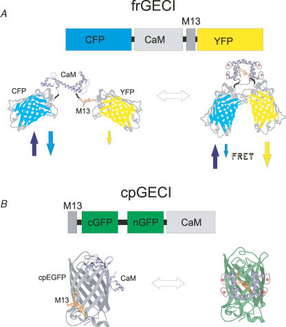
A, FRET-based genetically encoded Ca2+ indicators (frGECIs) exploit the conformational change associated with binding of Ca2+ to a Ca2+-binding molecule. The yellow cameleon model using calmodulin (CaM) and the 13-residue peptide-binding domain of myosin light chain kinase (M13) is shown above. Below, condensation of Ca2+/CaM/M13 reduces the separation between the donor, cyan fluorescent protein (CFP), and the acceptor, yellow fluorescent protein (YFP). Red balls represent Ca2+ ions in EF hands; arrows represent excitation (up arrow) and emission. B, circularly permutated indicators (cpGECIs) attach the Ca2+-binding protein and the target peptide to opposing ends of one of the β sheets forming the cage around the chromophore. Binding of Ca2+ results in an interaction between CaM and M13, rearranging the cage and enabling fluorescence (represented by green hue). cGFP and nGFP refer to the original C and N-terminal sequences.
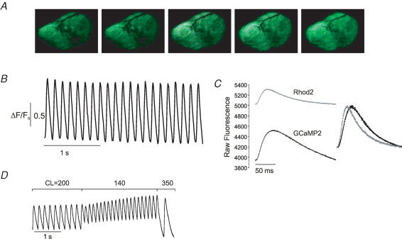
A, sequential in vivo images of the left ventricle of an anaesthetized, open-chested transgenic mouse expressing GCaMP2 demonstrate the signal change between end-diastole (left) and peak systole (right). Unprocessed colour images were taken with a consumer grade digital videocamera through a binocular fluorescence dissecting microscope (Leica MZFLIII). B, in vivo fluorescence transients from a 4 pixel area in the left atrium. C, fluorescence transients taken from an isolated, perfused GCaMP2 heart loaded with Rhod2. Note higher ΔF and lower background fluorescence in GCaMP2 signal. Overlay of the normalized traces demonstrates the underestimation of the rate of rise and decay of the Ca2+ signal by GCaMP2 in uncalibrated signals. D, pacing of a GCaMP2 heart in vitro demonstrates the consequence of GCaMP2 transition kinetics for rapid physiological signals. CL = cycle length.
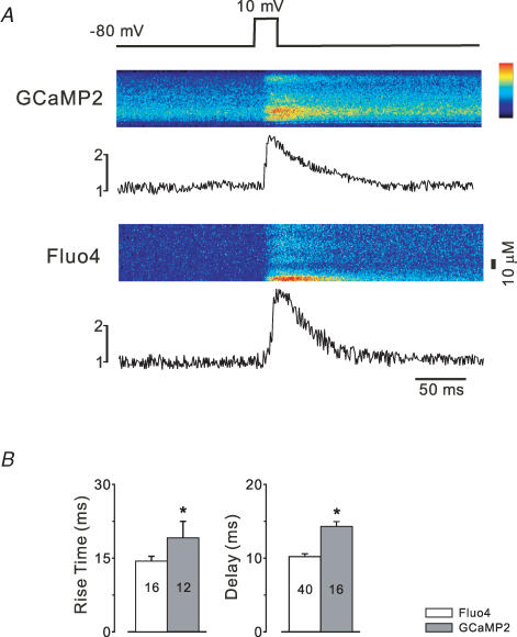
A, ventricular myocytes disaggregated from GCaMP2-expressing hearts were step-depolarized in voltage-clamp experiments and compared with myocytes from non-transgenic littermates loaded with Fluo4. Both preparations showed rapid fluorescence responses following step depolarization; however, the GCaMP2 signal decayed at a slower rate. B, the rise time and delay to rise was significantly longer in GECI-expressing cells.
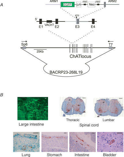
A, targeting strategy used to homologously transfer the EGFP coding sequence into the initiation codon of the choline acetyltransferase (ChAT) locus contained within a bacterial artificial chromosome. Note that the vesicular choline acetyltransferase (VAChT) gene is contained within the first intron of ChAT. B, expression of EGFP in BAC transgenic mice demonstrates high expression within the central and peripheral nervous systems, with precise marking of cholinergic enteric neurons. Scale bars are 100 μm (large intestine), 250 μm (spinal cord), and 50 μm (other). Adapted from Tallini et al. (2006b).
References
-
- Adams SR, Harootunian AT, Buechler YJ, Taylor SS, Tsien RY. Fluorescence ratio imaging of cyclic AMP in single cells. Nature. 1991;349:694–697. - PubMed
-
- Bacskai BJ, Hochner B, Mahaut-Smith M, Adams SR, Kaang BK, Kandel ER, Tsien RY. Spatially resolved dynamics of cAMP and protein kinase A subunits in Aplysia sensory neurons. Science. 1993;260:222–226. - PubMed
-
- Baker LC, London B, Choi BR, Koren G, Salama G. Enhanced dispersion of repolarization and refractoriness in transgenic mouse hearts promotes reentrant ventricular tachycardia. Circ Res. 2000;86:396–407. - PubMed
-
- Benaim G, Villalobo A. Phosphorylation of calmodulin. Functional implications. Eur J Biochem. 2002;269:3619–3631. - PubMed
Publication types
MeSH terms
Substances
Grants and funding
LinkOut - more resources
Full Text Sources
Other Literature Sources
Miscellaneous