. 2006 Dec 15;577(Pt 3):815-28.
doi: 10.1113/jphysiol.2006.117648. Epub 2006 Oct 12.

Mechanosensitive currents in the neurites of cultured mouse sensory neurones

Affiliations

Mechanosensitive currents in the neurites of cultured mouse sensory neurones

Jing Hu et al. J Physiol. .

Abstract

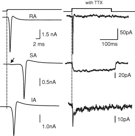
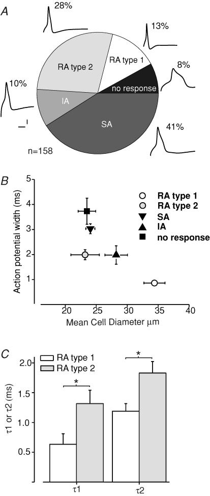
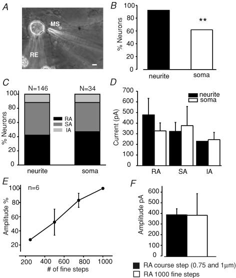
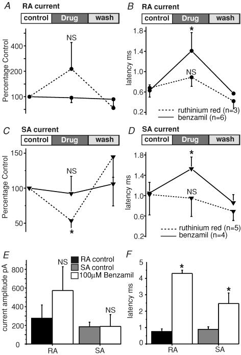
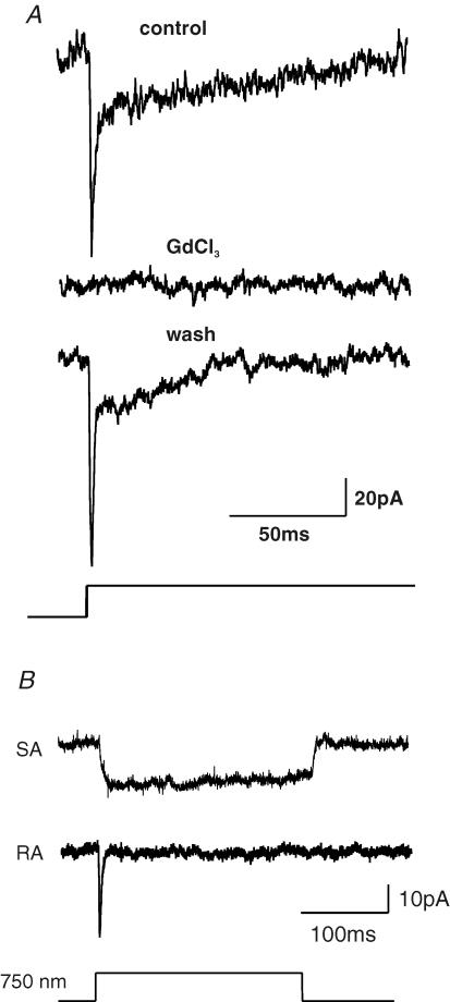
Almost all sensory neurones in the dorsal root ganglia have a mechanosensory function. The transduction of mechanical stimuli in vivo takes place exclusively at the sensory ending. For cutaneous sensory receptors it has so far proved impossible to directly record the mechanically gated receptor potential because of the small size and inaccessibility of the sensory ending. Here we investigate whether mechanosensitive currents are present in the neurites of freshly isolated adult mouse sensory neurones in culture. Almost all sensory neurone neurites possess currents gated by submicrometre displacement stimuli (92%). Three types of mechanically activated conductance were characterized based on different inactivation kinetics. A rapidly adapting conductance was found in larger sensory neurones with narrow action potentials characteristic of mechanoreceptors. Slowly and intermediate adapting conductances were found exclusively in putative nociceptive neurones. Mechanically activated currents with similar kinetics were found also after stimulating the cell soma. However, soma currents were only observed in around 60% of cells tested and the displacement threshold was several times larger than for the neurite (approximately 6 microm). The reversal potential of the rapidly adapting current indicated that this current is largely selective for sodium ions whereas the slowly adapting current is non-selective. It is likely that distinct ion channel entities underlie these two currents. In summary, our data suggest that the high sensitivity and robustness of mechanically gated currents in the sensory neurite make this a useful in vitro model for the mechanosensitive sensory endings in vivo.


Figures

Figure 1
Figure 1. Mechanosensitivity of DRG neurones cultured on poly-l-lysine–laminin substrate
A, images of single DRG neurone in the whole-cell recording configuration with the mechanical stimulator (MS) poised to stimulate one of the neurites. Phase-contrast image (top) and fluorescent image (bottom). The cell and neurites were loaded with Lucifer Yellow dye via the recording electrode (RE). The arrow shows the site of stimulation with the MS. Scale, 10 μm. B, sample traces of the different types of mechanically gated currents obtained. RA, rapidly adapting; SA, slowly adapting; IA, intermediate adapting. C, example of an RA current drawn on an expanded scale showing how mechanical latency was measured. D, the mean mechanical latency for RA, IA and SA currents are shown. The mean measured latency for the RA and SA current was not different but IA currents were activated with significantly shorter latencies than both RA and SA currents (*P < 0.01, unpaired t test).
Figure 2
Figure 2. Mechanically activated currents before and after application of TTX
Sample current traces on the left show the whole-cell recording in the absence of TTX for three cells. The membrane potential at the cell soma was clamped at −60 mV. In each case a large transient inward current with a variable latency was evoked by the mechanical stimulus that probably represents the arrival of an unclamped action potential at the cell soma. Note that each cell could subsequently be classified as possessing an RA, SA or IA current after the response was evoked in the presence of TTX (right). Note that the amplitude and kinetics of the mechanically activated currents differ significantly from those observed in the absence of TTX (1 μm). Note that for the neurone that possessed an SA current (middle traces), the beginning of inward current activation can be seen to precede the action potential when recorded in the absence of TTX (arrow).
Figure 3
Figure 3. Expression of the mechanically activated current in mechanoreceptors and nociceptors
A, the distribution of mechanically gated currents in the total population of neurones studied (n = 158) is shown in a pie chart. We have subdivided neurones with an RA current into type 1 and type 2 cells that are chiefly distinguished by the absence or presence of a hump on the falling phase of the action potential. Example traces of the measured action potential configuration for each cell type are shown together with the percentage of the total population adjacent to each pie chart slice. Scale bar for action potential correspond to 2 ms and 10 mV. B, the action potential width (measured at 50% amplitude) is plotted against the soma diameter for cells exhibiting RA, SA, IA or no mechanically gated current. Note that type 1 and type 2 cells with RA currents have very different cell sizes and action potentials widths (P < 0.001 unpaired t test for both parametres). RA type 2 and cells with IA currents had similar action potential widths but IA cells were significantly larger than RA type 2 cells (P < 0.05 unpaired t test). Cells with an SA current had significantly broader action potentials than RA type 1, RA type 2 and IA cells (P < 0.001 unpaired t test). C, the activation time constant τ1 and the inactivation time constant τ2 are plotted for RA type 1 and type 2 cells separately. It was clear that the RA type 1 cells have significantly faster activation and inactivation kinetics than type 2 cells (*P < 0.02 unpaired t test).
Figure 4
Figure 4. Comparison of mechanically evoked currents in the soma and neurite
A, the photomicrograph shows an example of the recording and stimulation procedure for evoking somal currents. Scale, 10 μm. B, the proportion of cells in which any mechanically evoked current could be evoked in the soma was significantly smaller (P < 0.001, χ2 test) than that found in the neurite. C, when we analysed the proportion of only those cells with a mechanosensitive current, the incidence of the RA, SA and IA current was not different. D, the mean amplitude of the mechanically gated currents evoked from the soma was also not significantly different from that found in the neurite. The data plotted are only from cells that were tested with a mechanical stimulus at the neurite and at the soma (n = 55). The amplitude of the displacement stimulus needed to evoke a somal current was many times larger than that used for neurites (see text). E, in some cells we also used smaller displacement stimuli (range, ∼250–1000 nm) applied to the neurite using the fine mode of the nanomotor. An analysis of six cells with an RA type current revealed that increasing displacement starting from 250 nm produced increasing current amplitudes in the same cells. F, for supratheshold stimuli the mean magnitude of the RA current was found to be the same regardless of whether the fine or coarse mode was used.
Figure 5
Figure 5. Heterogeneity of mechanically gated currents in sensory neurites
A, current–voltage relations for the mechanically activated current evoked by stimulating the cell soma or neurite immediately adjacent to the soma. In the case of cells with an RA current (n = 13), a linear fit of the data indicated that the current reverses at around +80 mV. Note both RA type 1 and type 2 cells were included in this analysis and no difference was seen in their IV relation. B, in contrast, for data obtained from cells with an SA response (n = 8), a linear fit indicated reversal at or slightly positive to 0 mV. C, replacement of sodium ions in the extracellular solution by the non-permeant cation NMDG+ completely blocked the RA current (n = 10) measured at −60 mV. No significant effect of NMDG+ ions was observed on the SA current (n = 3).
Figure 6
Figure 6. Differential pharmacological sensitivity of SA and RA currents in sensory neurites
A, cells with an RA current were incubated with 10 μm ruthenium red (n = 3) or 100 μm benzamil (n = 6) and the current amplitude measured before, during and after drug application. No significant effect of ruthenium red on the current amplitude was noted. B, the kinetic parameters of the RA current was also monitored during drug application and a significant increase in the latency for current activation was found in the presence of benzamil but not ruthenium red. C, in contrast to the RA current the mean amplitude of the SA current could be partially blocked by 10 μm ruthenium red (n = 5); 100 μm benzamil again had no significant effect on the current (n = 4). D, benzamil but not ruthenium red caused a significant and reversible increase in the latency for SA current activation. E, mechanically gated currents were measured in the continued presence of 100 μm benazamil. The amplitude of SA (n = 9) and RA (n = 9) currents were not significantly affected by the presence of benzamil. F, the latency for current activation was dramatically changed in the presence of benzamil with latency increasing more than 5-fold in the case of RA cells (*P < 0,05 unpaired t test).
Figure 7
Figure 7. Sample traces illustrating reversible blockade of the mechanosensitive current by gadolinium and presence of RA and SA currents in single cells
A, sample traces before, during and after incubation of the cell with gadolinium (10 μm). Note that the current is completely and reversible blocked by gadolimium. B, example traces recorded by stimulating the neuritic tree of a single sensory neurone at two locations. In rare cases, such as illustrated, an SA or RA current were evoked at the two locations.

Similar articles

Cited by

References

    1. Akopian AN, Sivilotti L, Wood JN. A tetrodotoxin-resistant voltage-gated sodium channel expressed by sensory neurons. Nature. 1996;379:257–262. - PubMed
    1. Cho H, Koo JY, Kim S, Park SP, Yang Y, Oh U. A novel mechanosensitive channel identified in sensory neurons. Eur J Neurosci. 2006;23:2543–2550. - PubMed
    1. Cho H, Shin J, Shin CY, Lee SY, Oh U. Mechanosensitive ion channels in cultured sensory neurons of neonatal rats. J Neurosci. 2002;22:1238–1247. - PMC - PubMed
    1. Clapham DE, Julius D, Montell C, Schultz G. International Union of Pharmacology. XLIX. Nomenclature and structure-function relationships of transient receptor potential channels. Pharmacol Rev. 2005;57:427–450. - PubMed
    1. Cunningham JT, Wachtel RE, Abboud FM. Mechanosensitive currents in putative aortic baroreceptor neurons in vitro. J Neurophysiol. 1995;73:2094–2098. - PubMed

Publication types

Substances

LinkOut - more resources