. 2006 Oct 24;103(43):15951-6.
doi: 10.1073/pnas.0604493103. Epub 2006 Oct 12.

Disseminated and sustained HIV infection in CD34+ cord blood cell-transplanted Rag2-/-gamma c-/- mice

Affiliations

Disseminated and sustained HIV infection in CD34+ cord blood cell-transplanted Rag2-/-gamma c-/- mice

Stefan Baenziger et al. Proc Natl Acad Sci U S A. .

Abstract

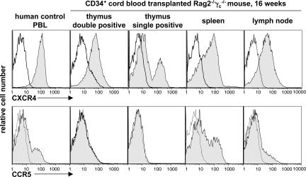
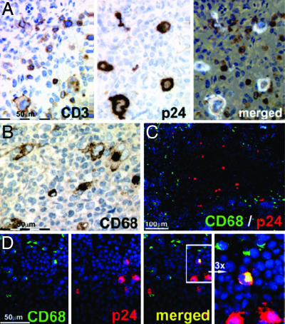
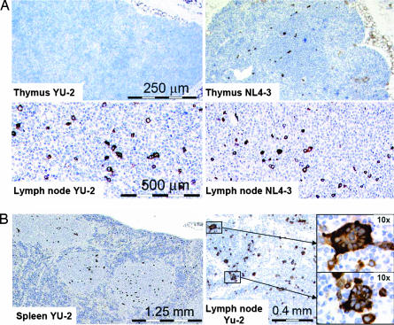
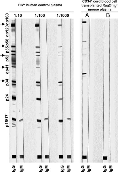
Because of species selectivity, HIV research is largely restricted to in vitro or clinical studies, both limited in their ability to rapidly assess new strategies to fight the virus. To prospectively study some aspects of HIV in vivo, immunodeficient mice, transplanted with either human peripheral blood leukocytes or human fetal tissues, have been developed. Although these are susceptible to HIV infection, xenoreactivity, and short infection spans, resource and ethical constraints, as well as biased HIV coreceptor tropic strain infection, pose substantial problems in their use. Rag2(-/-)gamma(c)(-/-) mice, transplanted as newborns with human CD34(+) cells, were recently shown to develop human B, T, and dendritic cells, constituting lymphoid organs in situ. Here we tested these mice as a model system for HIV-1 infection. HIV RNA levels peaked to up to 2 x 10(6) copies per milliliter of plasma early after infection, and viremia was observed for up to 190 days, the longest time followed. A marked relative CD4(+) T cell depletion in peripheral blood occurred in CXCR4-tropic strain-infected mice, whereas this was less pronounced in CCR5-tropic strain-infected animals. Thymus infection was almost exclusively observed in CXCR4-tropic strain-infected mice, whereas spleen and lymph node HIV infection occurred irrespective of coreceptor selectivity, consistent with respective coreceptor expression on human CD4(+) T cells. Thus, this straightforward to generate and cost-effective in vivo model closely resembles HIV infection in man and therefore should be valuable to study virus-induced pathology and to rapidly evaluate new approaches aiming to prevent or treat HIV infection.


Conflict of interest statement

The authors declare no conflict of interest.

Figures

Fig. 1.
Fig. 1.
CCR5 and CXCR4 expression on human CD4+ cells in CD34+ cell-transplanted Rag2−/−γc−/− mice resembles expression patterns in humans. Histograms depict representative receptor expressions (shaded histograms) and respective isotype controls (open histograms) on CD4+ gated control human PBL and cells isolated from tissue of a mouse at 16 weeks after birth and transplantation of CD34+ cells.
Fig. 2.
Fig. 2.
Long-term and high-titer CCR5- and CXCR4-tropic HIV infection in human CD34+ cord blood cell-transplanted Rag2−/−γc−/− mice. (A) Quantitative HIV RNA plasma levels in animals successfully infected with CCR5-tropic (YU-2; n = 13) and CXCR4-tropic (NL4-3; n = 18) HIV strains. Black triangles and connector lines indicate sequential analysis of single mice, and brown diamonds indicate mice analyzed at a single time point. (B) Graphs depict quantitative HIV RNA plasma levels (copies per milliliter of plasma; right y axis) and relative CD4+ and CD8+ T cell levels (percentage of human CD45+ blood cells; left y axis) in individual mice over time (uninfected, n = 2; YU-2- and NL4-3-infected, n = 4 each), showing more pronounced CD4+ T cell depletion in CXCR4-tropic infected animals.
Fig. 3.
Fig. 3.
HIV p24+ cells are mostly human CD3+ cells and only occasionally non-T cells such as CD68+ macrophages. (A) Histologies show consecutive spleen sections (paraffin, 1 μm) stained with antibodies against human CD3 (Left) and p24 (Center) and a merged presentation of both (Right) in a YU-2-infected animal 18 days after infection. (B) Anti-human CD68 staining on paraffin-embedded material. (C) Merged anti-CD68 (green), HIV-p24 (red), and DAPI (blue) staining of cryoembedded spleen (6-μm sections) showing that p24+ cells mainly localize in white pulp areas (darker area; see also Fig. 4B), whereas CD68+ cells mostly localize at adjacent margins and red pulp areas. (D) Consecutive spleen cryosection staining and respective merged presentation showing a rare CD68 and p24 double-positive cell (yellow). The far-right image shows a ×3 enlargement of the area with the double-positive cell. (B–D) Representative spleen sections from a YU-2-infected animal 23 days after infection.
Fig. 4.
Fig. 4.
HIV spreading in lymphoid organs of human CD34+ cell-transplanted Rag2−/−γc−/− mice resembles HIV infection in humans. (A) Representative p24-stained thymus and lymph node sections of YU-2- and NL4-3-infected mice analyzed at 37 and 23 days after infection, respectively. No or very rare p24 staining is observed in thymi of CCR5-tropic YU-2-infected animals. (B) Representative tissue section of spleen (Left) and lymph node (Right) of a YU-2-infected animal at 52 days after infection showing multinucleated p24+ giant cells (enlarged Insets Right).
Fig. 5.
Fig. 5.
Humoral immune response in HIV-infected human CD34+ cell-transplanted Rag2−/−γc−/− mice. Western blot analysis of HIV-specific IgG and IgM responses is shown. (Left) Three-step dilution of plasma from an HIV-infected patient (no antiviral therapy) showing full IgG seroconversion. (Right) Undiluted plasma of an animal showing a measurable IgG response against p34, gp41, p52, p58, and gp160 (lane A) and undiluted plasma of an animal showing no response (lane B).

References

    1. Jamieson BD, Zack JA. AIDS. 1999;13(Suppl A):S5–S11. - PubMed
    1. Levy JA. J Med Primatol. 1996;25:163–174. - PubMed
    1. Nath BM, Schumann KE, Boyer JD. Trends Microbiol. 2000;8:426–431. - PubMed
    1. Joag SV. Microbes Infect. 2000;2:223–229. - PubMed
    1. VandeBerg JL, Zola SM. Nature. 2005;437:30–32. - PubMed

Publication types

MeSH terms