. 2007 Feb;25(2):411-8.
doi: 10.1634/stemcells.2006-0380. Epub 2006 Oct 12.

Enhanced yield of neuroepithelial precursors and midbrain-like dopaminergic neurons from human embryonic stem cells using the bone morphogenic protein antagonist noggin

Affiliations

Enhanced yield of neuroepithelial precursors and midbrain-like dopaminergic neurons from human embryonic stem cells using the bone morphogenic protein antagonist noggin

Kai-Christian Sonntag et al. Stem Cells. 2007 Feb.

Abstract

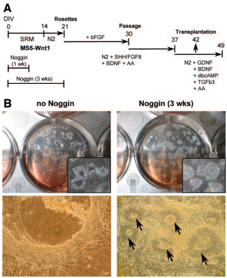
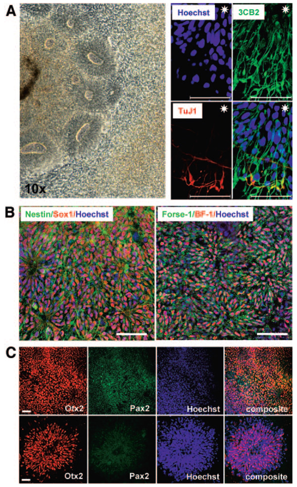
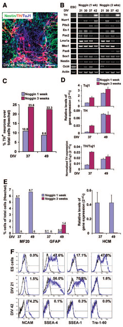
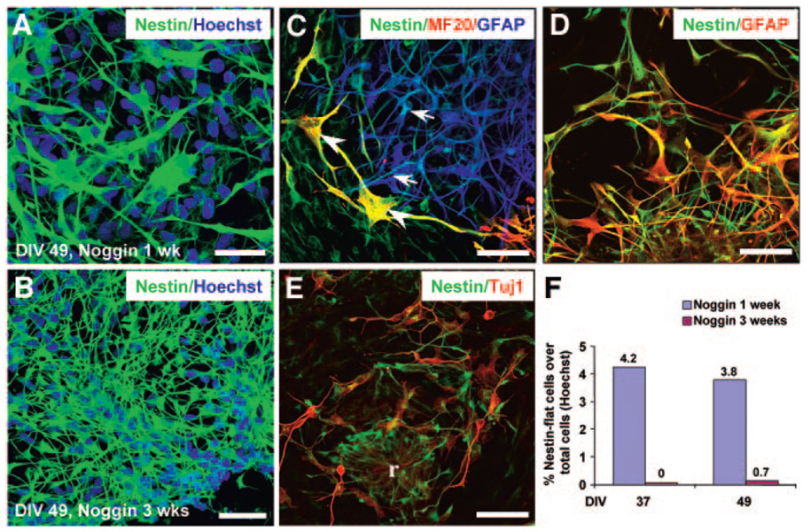
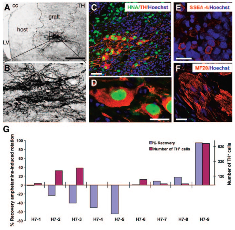
It is currently not known whether dopamine (DA) neurons derived from human embryonic stem cells (hESCs) can survive in vivo and alleviate symptoms in models of Parkinson disease (PD). Here, we report the use of Noggin (a bone morphogenic protein antagonist) to induce neuroectodermal cell development and increase the yield of DA neurons from hESCs. A combination of stromal-derived inducing activity and Noggin markedly enhanced the generation of neuroepithelial progenitors that could give rise to DA neurons. In addition, Noggin diminished the occurrence of a fibroblast-like Nestin-positive precursor population that differentiated into myocytes. After transplantation of differentiated hESCs to a rodent model of PD, some grafts contained human midbrain-like DA neurons. This protocol demonstrates hESC derivation and survival of human DA neurons appropriate for cell therapy in PD.


Conflict of interest statement

DISCLOSURES

The authors indicate no potential conflicts of interest.

Figures

Figure 1
Figure 1
Culture conditions and development of neuroectodermal cells. (A): Schematic representation of the protocol used to differentiate human embryonic stem cells (hESCs). hESCs were differentiated in a multistep protocol supporting dopamine neuronal cell development [4] and modified by adding 300 ng/ml Noggin during the stromal feeder cell-based neuroectodermal induction (DIV0–21) for either 1 week (DIV0–7) or 3 weeks (DIV 0–21). On DIV21, rosettes were selected and manually transferred to polyornithine/laminin-coated culture dishes, expanded, and further differentiated using the sequential addition of media supplements as indicated (see Material and Methods for further details). At DIV42, cells were harvested for transplantation into the striatum of 6-hydroxydopamine-treated rats. (B): The effects of Noggin on neuroectodermal cell development. Left panels: In the absence of Noggin, hESC colonies developed into typical crater-like structures with a single layer of homogeneous cells in the center and a rim with heterogeneous cellular morphologies. In these conditions, the colonies had very little to no rosettes. An overview of colonies at low magnification and a representation of four colonies at higher magnification are shown in the inset (×5). Right panels: Addition of Noggin for 3-weeks led to increased formation of rosette-forming ESC colonies. An overview of colonies from Noggin-treated cultures at low magnification, which macroscopically appear homogeneous with a high cell density, is shown. Bright-field image (×5) of the border of a single colony at DIV21 showing an accumulation of rosettes (arrows). Abbreviations: AA, ascorbic acid; BDNF, brain-derived neurotrophic factor; bFGF, basic fibroblast growth factor; DIV, day in vitro; FGF8, fibroblast growth factor 8; SHH, sonic hedgehog; SRM, serum replacement medium; TGF-β3, transforming growth factor type β3.
Figure 2
Figure 2
Characterization of the neuroectodermal precursors. (A): Left panel: Bright-field image (×10) of cultured rosettes at day in vitro 30 (DIV30). Well-defined rosettes were surrounded by proliferating precursors. Right panel: Immunocytochemistry (ICC) images of rosettes using the nuclear marker Hoechst (blue), the radial-glia marker 3CB2 (green), and the neuronal marker Tuj1 (red). Shown is a part of a rosette with the center marked by a white star. Rosettes contained radial-glia-like cells in the center part and neurons at their edges. Scale bars = 50 µm. (B): ICC for the immature neural markers Sox1 (red) and Nestin (green). Rosettes were also stained with the forebrain-midbrain markers Otx2 (red), Pax2 (green) (C), and the forebrain markers FORSE-1 (green) and BF-1 (red) at DIV21. (C): In all culture conditions (Noggin for 1 week or 3 weeks), colonies were either Otx2+/Pax2+ or Otx+/Pax2. Scale bars = 75 µm (left set of images), 40 µm (right set of images). Abbreviations: BF-1, brain factor-1; FORSE-1, forebrain surface embryonic marker 1.
Figure 3
Figure 3
The effect of Noggin on the development of neural and non-neural cellular phenotypes. (A): Dopamine (DA) neuronal cell development. Immunocytochemistry for Nestin (green), the neuronal marker Tuj1 (blue), the DA marker TH (red), and Hoechst at DIV49 in the 3-week Noggin conditions. Scale bars = 50 µm. (B): Gene expression profile during human embryonic stem cell (hESC) development. Reverse transcription-polymerase chain reaction (PCR) results from immature ESCs and cell samples at DIVs 21, 30, 37, and 42 for the 1-week or 3-week Noggin conditions. Samples were analyzed for markers related to the midbrain DA neuronal phenotype (TH, Nurr1, Pitx3, En-1, Pax2, Lmx1a, and Msx-1), the anterior segment marker Pax6, the neural precursor markers Sox1 and Nestin, and the immature ESC marker Oct4. Shown is one representative out of three independent experiments. (C): Quantification of the TH+ neurons at DIV37 and 49 in the Noggin 1-week (blue bars) and 3-week (red bars) conditions. Shown is one out of two experiments representing randomized cell counts of 4,000–8,000 Hoechst+ cells for each experiment. The numbers of TH+ cells are plotted as percentage of total cell counts as determined by Hoechst staining. (D): Quantitative real-time (Q)-PCR for Tuj1 and TH gene expression at DIV37 and 49. Relative gene expression levels were calculated using the 2−ΔCt method according to Livak et al. [20] (upper and middle panels). In addition, the TH was normalized to the corresponding Tuj1 expression (lower panel). (E): Quantification of the MF20+ and GFAP+ cell populations at DIV37 and 49 in the Noggin 1-week (blue bars) and 3-week (red bars) conditions as described in (B) (left panel). Right panel: Q-PCR for human heavy chain myosin (HCM) plotted as relative levels of gene expression (2−ΔCt) according to Livak et al. [20] (F): Detection of immature stem cells during hESC development in the 3-week Noggin condition. Fluorescence-activated cell sorting using specific antibodies for the stem cell markers SSEA-1, SSEA-4, and Tra-1-60, and the adhesion molecule NCAM at the immature ESC stage, DIV21 and 42. The numbers of immature ESCs substantially decreased during in vitro differentiation, whereas the fraction of NCAM+ cells increased. Abbreviations: Ct, threshold cycle; DIV, day in vitro; ESC, embryonic stem cell; GFAP, glial fibrillary acidic protein; MF20, myosin; NCAM, neural cell adhesion molecule; TH, tyrosine hydroxylase; Tuj1, β-III-tubulin.
Figure 4
Figure 4
The effects of Noggin on immature Nestin+ progenitor cell development. Characterization of Nestin+ cellular phenotypes at DIV49 in the 1-week (A) and 3-week (B) Noggin cultures. Scale bars = 50 µm. “Nestin-slender” cells were dominant and present in both culture conditions, whereas the “Nestin-flat” cells did not appear before DIV37 and occurred predominantly in the 1-week Noggin treatment condition. (C–E): Phenotype determination of the two Nestin+ cell populations shows that Nestin-flat cells coexpress MF20 (arrow-heads) (C) and that some Nestin-slender cells coexpress Tuj1 (E). GFAP is expressed in Nestin-flat cell populations (D) and arrows in (C). Scale bars = 50 µm. (F): Quantification of the Nestin-flat cell populations at DIV37 and 49 for the 1-week and 3-week Noggin cultures. Cell counts were performed as described in the legend for Figure 3C and 3E. Abbreviations: DIV, day in vitro; GFAP, glial fibrillary acidic protein; MF20, myosin; Tuj1, β-III-tubulin.
Figure 5
Figure 5
Typical cellular phenotypes in grafts after transplantation of differentiated human embryonic stem cells (hESCs) (here, H7). (A–F): Immunocytochemistry revealed numerous surviving TH+ neurons, sometimes in clusters, within the grafts in the 6-hydroxydopamine-lesioned rat striatum. These neurons had a mature morphology with big, polygonal cell soma (25–35 µm) and complex neuritic arborization. Scale bars = 1,000 µm (A), 50 µm (B). The TH+ neurons were derived from hESCs as demonstrated by coexpression of human nuclear antigen (HNA) (green) and TH (red). Hoechst nuclear counterstain is shown in blue. Scale bars = 50 µm (C), 25 µm (D). The grafts also contained cells that stained with SSEA-4-specific antibodies (red), indicating the presence of pluripotent immature precursors (E). Scale bar = 20 µm. (F): Example of non-neural multinucleated muscle cells within the graft (MF20 in red and Hoechst nuclear counterstain in blue). Scale bar = 40 µm. (G): Graphical representation of behavioral improvement in amphetamine-induced rotation (blue bars) paired with numbers of TH+ neurons within grafts (red bars) of H7 hESC (n = 9) transplanted animals 12 weeks after implantation. Each set of bars represents data from one animal (H7-1 to H7-9). Abbreviations: cc, corpus callosum; LV, left ventricle; MF20, myosin; TH, tyrosine hydroxylase.

References

    1. Zeng X, Cai J, Chen J, et al. Dopaminergic differentiation of human embryonic stem cells. STEM CELLS. 2004;22:925–940. - PubMed
    1. Buytaert-Hoefen KA, Alvarez E, Freed CR. Generation of tyrosine hydroxylase positive neurons from human embryonic stem cells after coculture with cellular substrates and exposure to GDNF. STEM CELLS. 2004;22:669–674. - PubMed
    1. Ben-Hur T, Idelson M, Khaner H, et al. Transplantation of human embryonic stem cell-derived neural progenitors improves behavioral deficit in Parkinsonian rats. STEM CELLS. 2004;22:1246–1255. - PubMed
    1. Perrier AL, Tabar V, Barberi T, et al. Derivation of midbrain dopamine neurons from human embryonic stem cells. Proc Natl Acad Sci U S A. 2004;101:12543–12548. - PMC - PubMed
    1. Park CH, Minn YK, Lee JY, et al. In vitro and in vivo analyses of human embryonic stem cell-derived dopamine neurons. J Neurochem. 2005;92:1265–1276. - PubMed

Publication types

MeSH terms