. 2006 Nov;7(11):1162-7.
doi: 10.1038/sj.embor.7400826. Epub 2006 Oct 13.

The neurite outgrowth inhibitor Nogo-A promotes denervation in an amyotrophic lateral sclerosis model

Affiliations

The neurite outgrowth inhibitor Nogo-A promotes denervation in an amyotrophic lateral sclerosis model

Natasa Jokic et al. EMBO Rep. 2006 Nov.

Abstract

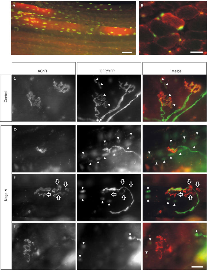
Amyotrophic lateral sclerosis (ALS) is a neurodegenerative disease characterized by motor neuron loss and muscle wasting. In muscles of ALS patients, Nogo-A-a protein known to inhibit axon regeneration-is ectopically expressed at levels that correlate with the severity of the clinical symptoms. We now show that the genetic ablation of Nogo-A extends survival and reduces muscle denervation in a mouse model of ALS. In turn, overexpression of Nogo-A in wild-type muscle fibres leads to shrinkage of the postsynapse and retraction of the presynaptic motor ending. This suggests that the expression of Nogo-A occurring early in ALS skeletal muscle could cause repulsion and destabilization of the motor nerve terminals, and subsequent dying back of the axons and motor neurons.


Figures

Figure 1
Figure 1
Genetic ablation of Nogo-A in Sod1(G86R) mice. (A) Cumulative probability of survival of G86R/Nogo-A+/+ and G86R/Nogo-A−/− mice (P=0.04, n=16–17 mice, log-rank test). (B) Cell counts in the ventral horns of the lumbar spinal cord of G86R/Nogo-A+/+ and G86R/Nogo-A−/− mice. Toluidine-blue-stained cells were divided into two groups of 300–600μm2 and >600 μm2, the latter clearly representing motor neurons. No differences were observed in the number of cells between non-Cu/Zn-superoxide dismutase 1 (Sod1)(G86R) Nogo-A-knockout and wild-type mice (data not shown; *P<0.05 versus corresponding +/+, n=3 mice, Student's t-test). (C) Ubiquitin immunoreactivity in large motor neurons (cross-sectional area >600 μm2; arrowheads) of the lumbar spinal cord from G86R/Nogo-A+/+ and G86R/Nogo-A−/− mice. Scale bar, 30 μm. (D) Messenger RNA levels of acetylcholine receptor α-subunit (AChRα) and muscle-specific receptor tyrosine kinase (MuSK) in the soleus muscle of wild-type (WT; open bars) and Sod1(G86R) mice (filled bars) containing (+/+) or not containing (−/−) Nogo-A (***P<0.001 versus wild type, #P<0.05 versus +/+, n=4–5 mice, analysis of variance followed by Tukey's test). (E) Levels of MuSK protein as determined by western blot of immunoprecipitated MuSK from muscle extracts of Sod1(G86R) mice containing (+/+) or not containing (−/−) Nogo-A. MW, molecular weight markers. In all cases, symptomatic animals reached the end point of the disease. Sod1, Cu/Zn-superoxide dismutase 1.
Figure 2
Figure 2
Overexpression of Nogo-A in muscle causes the disassembly of the neuromuscular junction. Electroporation of the soleus muscle with expression constructs for Nogo-A and nuclear-localized green fluorescent protein (GFP) results in muscle fibres that express high levels of Nogo-A (red) and GFP (green) after 2 weeks. (A) Whole-mount and (B) cross-section of the soleus muscle. Representative pictures of neuromuscular junctions (NMJs), 6 weeks after electroporation of empty vector (control; C) or Nogo-A expression constructs (DF). The postsynaptic apparatus was visualized by rhodamine-labelled α-bungarotoxin (acetylcholine receptor; red in merge). Presynaptic nerve terminals were labelled by yellow fluorescent protein (YFP; green in merge) and transfected muscle fibres by nuclear localization signal–GFP (GFP, green in merge). For clarification, GFP-positive myonuclei are indicated by arrowheads. In control transfections, both pre- and postsynaptic structures look normal (C). By contrast, overexpression of Nogo-A often results in smaller NMJs (D) and fragmentation of the postsynaptic structure including partial (E; hollow arrows) or total (F; asterisks) retraction of the presynaptic nerve terminal. Scale bar, 25 μm.

References

    1. Azzouz M, Ralph GS, Storkebaum E, Walmsley LE, Mitrophanous KA, Kingsman SM, Carmeliet P, Mazarakis ND (2004) VEGF delivery with retrogradely transported lentivector prolongs survival in a mouse ALS model. Nature 429: 413–417 - PubMed
    1. Bezakova G, Ruegg MA (2003) New insights into the roles of agrin. Nat Rev Mol Cell Biol 4: 295–308 - PubMed
    1. Bruijn LI, Miller TM, Cleveland DW (2004) Unraveling the mechanisms involved in motor neuron degeneration in ALS. Annu Rev Neurosci 27: 723–749 - PubMed
    1. Caroni P, Schwab ME (1988) Antibody against myelin-associated inhibitor of neurite growth neutralizes nonpermissive substrate properties of CNS white matter. Neuron 1: 85–96 - PubMed
    1. Clement AM et al. (2003) Wild-type nonneuronal cells extend survival of SOD1 mutant motor neurons in ALS mice. Science 302: 113–117 - PubMed

Publication types