. 2006;7(10):R91.
doi: 10.1186/gb-2006-7-10-r91. Epub 2006 Oct 13.

Independent centromere formation in a capricious, gene-free domain of chromosome 13q21 in Old World monkeys and pigs

Affiliations

Independent centromere formation in a capricious, gene-free domain of chromosome 13q21 in Old World monkeys and pigs

Maria Francesca Cardone et al. Genome Biol. 2006.

Abstract

Background: Evolutionary centromere repositioning and human analphoid neocentromeres occurring in clinical cases are, very likely, two stages of the same phenomenon whose properties still remain substantially obscure. Chromosome 13 is the chromosome with the highest number of neocentromeres. We reconstructed the mammalian evolutionary history of this chromosome and characterized two human neocentromeres at 13q21, in search of information that could improve our understanding of the relationship between evolutionarily new centromeres, inactivated centromeres, and clinical neocentromeres.

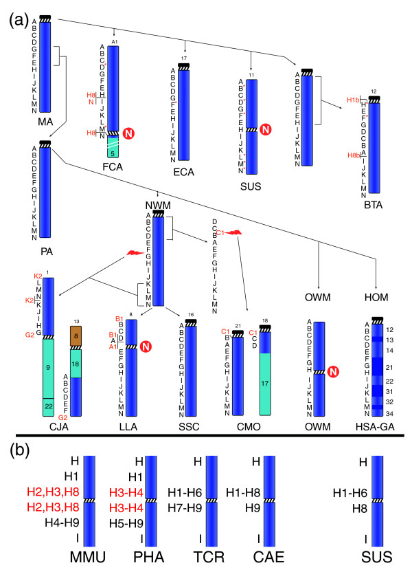
Results: Chromosome 13 evolution was studied, using FISH experiments, across several diverse superordinal phylogenetic clades spanning >100 million years of evolution. The analysis revealed exceptional conservation among primates (hominoids, Old World monkeys, and New World monkeys), Carnivora (cat), Perissodactyla (horse), and Cetartiodactyla (pig). In contrast, the centromeres in both Old World monkeys and pig have apparently repositioned independently to a central location (13q21). We compared these results to the positions of two human 13q21 neocentromeres using chromatin immunoprecipitation and genomic microarrays.

Conclusion: We show that a gene-desert region at 13q21 of approximately 3.9 Mb in size possesses an inherent potential to form evolutionarily new centromeres over, at least, approximately 95 million years of mammalian evolution. The striking absence of genes may represent an important property, making the region tolerant to the extensive pericentromeric reshuffling during subsequent evolution. Comparison of the pericentromeric organization of chromosome 13 in four Old World monkey species revealed many differences in sequence organization. The region contains clusters of duplicons showing peculiar features.


Figures

Figure 1
Figure 1
Examples of co-hybridization experiments on (a) macaque (MMU), (b) sacred baboon (PHA), (c) silvered-leaf monkey (TCR), (d) African green monkey (CAE), (e) pig (SUS), and (f) cat (FCA). The telomerically located probes in (a-e) were used for a correct identification of p and q arms of these metacentric chromosomes. The DAPI image alone is reported on the left to better show the morphology of the chromosome. Letters refer to BAC clones reported in Table 1.
Figure 2
Figure 2
Diagrammatic representation of the evolutionary history of chromosome 13. (a) Marker order arrangement in the studied species, from which the arrangement of the mammalian ancestor (MA) and primate ancestor (PA) was derived (see text). N in a red circle stands for new centromere. The number that identifies the chromosome in each species is reported on top of the chromosome. The black letters on the left of each primate chromosome refer to the panel of BAC probes reported in Table 1 (human BACs); letters on cattle (BTA), pig (SUS), horse (ECA), and cat (FCA) chromosomes refer to BACs reported in Additional data file 1, obtained by library screening or from published databases (see text). Letters in red are the additional probes used to delimit chromosomal breakpoints or featuring unusual results (see N and H8 in the cat). Letter with asterisk indicate BACs identified on the radiation hybrids mapping data and used to fill gaps due to library screenings failure (see Table 1 and text). The long arm of cat chromosome A1 was shortened because of space constraint. The red lightning indicates chromosome break. (b) Results of FISH experiments of the H1 to H9 clones (Table 1) on OWM species (left) and in pig (SUS, right). Clones in red are duplicated. In OWM, the clones not reported in the figure failed to yield FISH signals. For details see text. (HOM = Hominoidea;HSA-GA = Homo Sapiens-Great Apes Ancestor).
Figure 3
Figure 3
ChIP CHIP results on the two neocentromere cases. (a) Ideogrammatic representation of the neocentric chromosomes found in the two independent neocentromere cases. From left to right: the invdup13q14 chromosome with a neocentromere in band 13q21; the ring chromosome derived from band 13q21 to 13q22; a normal chromosome 13 for comparison. At the far right, the region 13q21.3 is expanded to show the relative position of BAC clones H2 to H9, along with the human neocentromeres and evolutionarily new centromeres of OWM and pig (see also Figure 2b). (b) ChIP on a CHIP analysis of the invdup13q14 chromosome using antibodies to CENP-A. (3) ChIP on a CHIP analysis of the ring13q21 chromosome using antibodies to CENP-C. For (b and c), the microarray was hybridized simultaneously with Cy-5 labeled CENP ChIP DNA (red) and Cy3 labeled input chromatin DNA (green). The scale normalized mean Log2 Cy-5:Cy-3 ratios and standard error for each BAC are shown plotted on the y-axis for three independent ChIP experiments. Alpha satellite DNA was included in the microarray as a positive control (far right). (3) Position of 107 contiguous BACs spanning 13q21 to 13q22 is shown across the x-axis. Positions of probes H7, H8, H9 and I (Table 1) are shown. Bottom: blow up of BACs that are positive for CENP proteins. Thin lines represent regions of BAC overlap. Scale in Mbp according to UCSC genome coordinates hg17 [Bioinformatics, #9821].

References

    1. du Sart D, Cancilla MR, Earle E, Mao J, Saffery R, Tainton KM, Kalitsis P, Martin J, Barry AE, Choo KHA. A functional neo centromere formed through activation of a latent human centromere and consisting of non-alpha-satellite DNA. Nat Genet. 1997;16:144–153. doi: 10.1038/ng0697-144. - DOI - PubMed
    1. Warburton PE. Epigenetic analysis of kinetochore assembly on variant human centromeres. Trends Genet. 2001;17:243–247. doi: 10.1016/S0168-9525(01)02283-1. - DOI - PubMed
    1. Amor DJ, Choo KH. Neocentromeres: role in human disease, evolution, and centromere study. Am J Hum Genet. 2002;71:695–714. doi: 10.1086/342730. - DOI - PMC - PubMed
    1. Warburton PE. Chromosomal dynamics of human neocentromere formation. Chromosome Res. 2004;12:617–626. doi: 10.1023/B:CHRO.0000036585.44138.4b. - DOI - PubMed
    1. Warburton PE, Dolled M, Mahmood R, Alonso A, Li S, Naritomi K, Tohma T, Nagai T, Hasegawa T, Ohashi H, et al. Molecular cytogenetic analysis of eight inversion duplications of human chromosome 13q that each contain a neocentromere. Am J Hum Genet. 2000;66:1794–1806. doi: 10.1086/302924. - DOI - PMC - PubMed

Publication types

LinkOut - more resources