

Frequency, prognostic impact, and subtype association of 8p12, 8q24, 11q13, 12p13, 17q12, and 20q13 amplifications in breast cancers
- PMID: 17040570
- PMCID: PMC1626089
- DOI: 10.1186/1471-2407-6-245
Frequency, prognostic impact, and subtype association of 8p12, 8q24, 11q13, 12p13, 17q12, and 20q13 amplifications in breast cancers
Abstract
Background: Oncogene amplification and overexpression occur in tumor cells. Amplification status may provide diagnostic and prognostic information and may lead to new treatment strategies. Chromosomal regions 8p12, 8q24, 11q13, 17q12 and 20q13 are recurrently amplified in breast cancers.
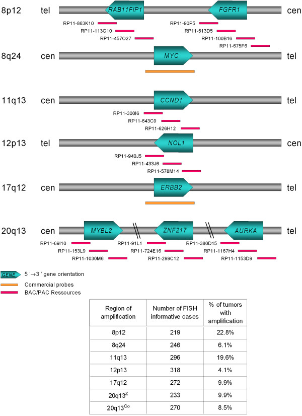
Methods: To assess the frequencies and clinical impact of amplifications, we analyzed 547 invasive breast tumors organized in a tissue microarray (TMA) by fluorescence in situ hybridization (FISH) and calculated correlations with histoclinical features and prognosis. BAC probes were designed for: (i) two 8p12 subregions centered on RAB11FIP1 and FGFR1 loci, respectively; (ii) 11q13 region centered on CCND1; (iii) 12p13 region spanning NOL1; and (iv) three 20q13 subregions centered on MYBL2, ZNF217 and AURKA, respectively. Regions 8q24 and 17q12 were analyzed with MYC and ERBB2 commercial probes, respectively.
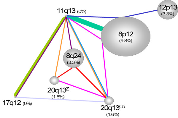
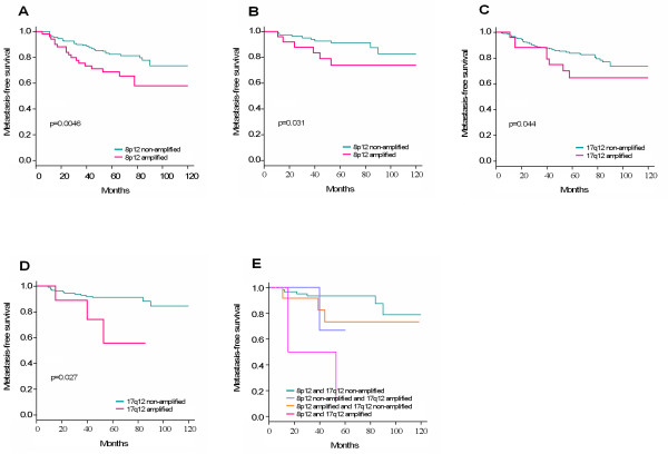
Results: We observed amplification of 8p12 (amplified at RAB11FIP1 and/or FGFR1) in 22.8%, 8q24 in 6.1%, 11q13 in 19.6%, 12p13 in 4.1%, 17q12 in 9.9%, 20q13Z (amplified at ZNF217 only) in 9.9%, and 20q13Co (co-amplification of two or three 20q13 loci) in 8.5% of cases. The 8q24, 12p13, and 17q12 amplifications were correlated with high grade. The most frequent single amplifications were 8p12 (9.8%), 8q24 (3.3%) and 12p13 (3.3%), 20q13Z and 20q13Co (1.6%) regions. The 17q12 and 11q13 regions were never found amplified alone. The most frequent co-amplification was 8p12/11q13. Amplifications of 8p12 and 17q12 were associated with poor outcome. Amplification of 12p13 was associated with basal molecular subtype.
Conclusion: Our results establish the frequencies, prognostic impacts and subtype associations of various amplifications and co-amplifications in breast cancers.
Figures
References
-
- Tanner MM, Tirkkonen M, Kallioniemi A, Isola J, Kuukasjarvi T, Collins C, Kowbel D, Guan XY, Trent J, Gray JW, Meltzer P, Kallioniemi OP. Independent amplification and frequent co-amplification of three nonsyntenic regions on the long arm of chromosome 20 in human breast cancer. Cancer Res. 1996;56:3441–3445. - PubMed
-
- Cuny M, Kramar A, Courjal F, Johannsdottir V, Iacopetta B, Fontaine H, Grenier J, Culine S, Theillet C. Relating genotype and phenotype in breast cancer: an analysis of the prognostic significance of amplification at eight different genes or loci and of p53 mutations. Cancer Res. 2000;60:1077–1083. - PubMed
-
- Al-Kuraya K, Schraml P, Torhorst J, Tapia C, Zaharieva B, Novotny H, Spichtin H, Maurer R, Mirlacher M, Kochli O, Zuber M, Dieterich H, Mross F, Wilber K, Simon R, Sauter G. Prognostic relevance of gene amplifications and coamplifications in breast cancer. Cancer Res. 2004;64:8534–8540. doi: 10.1158/0008-5472.CAN-04-1945. - DOI - PubMed
-
- Gelsi-Boyer V, Orsetti B, Cervera N, Finetti P, Sircoulomb F, Rouge C, Lasorsa L, Letessier A, Ginestier C, Monville F, Esteyries S, Adelaide J, Esterni B, Henry C, Ethier SP, Bibeau F, Mozziconacci MJ, Charafe-Jauffret E, Jacquemier J, Bertucci F, Birnbaum D, Theillet C, Chaffanet M. Comprehensive profiling of 8p11-12 amplification in breast cancer. Mol Cancer Res. 2005;3:655–667. doi: 10.1158/1541-7786.MCR-05-0128. - DOI - PubMed
Publication types
MeSH terms
Substances
LinkOut - more resources
Full Text Sources
Other Literature Sources
Medical
Molecular Biology Databases
Research Materials
Miscellaneous
