. 2006;34(20):5764-77.
doi: 10.1093/nar/gkl722. Epub 2006 Oct 13.

Characterization of a nucleocapsid-like region and of two distinct primer tRNALys,2 binding sites in the endogenous retrovirus Gypsy

Affiliations

Characterization of a nucleocapsid-like region and of two distinct primer tRNALys,2 binding sites in the endogenous retrovirus Gypsy

Caroline Gabus et al. Nucleic Acids Res. 2006.

Abstract

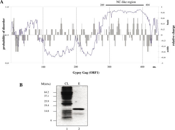
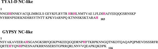
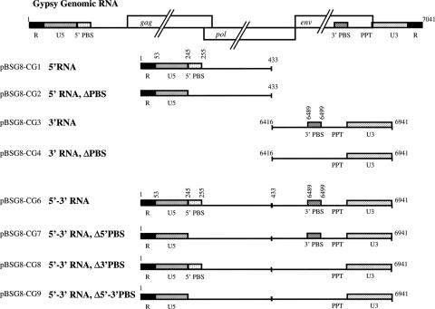
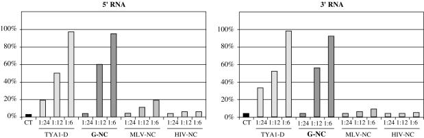
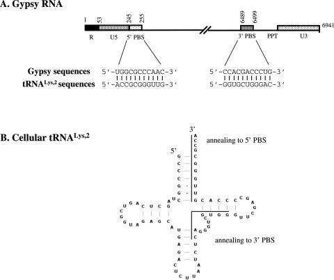
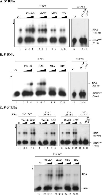
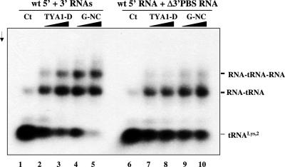
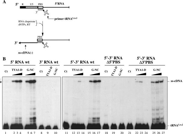
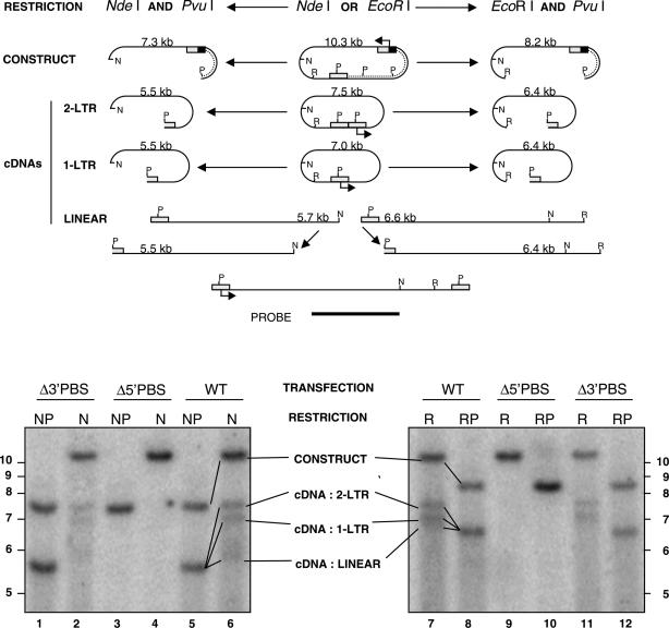
Mobile LTR-retroelements comprising retroviruses and LTR-retrotransposons form a large part of eukaryotic genomes. Their mode of replication and abundance favour the notion that they are major actors in eukaryote evolution. The Gypsy retroelement can spread in the germ line of the fruit fly Drosophila melanogaster via both env-independent and env-dependent processes. Thus, Gypsy is both an active retrotransposon and an infectious retrovirus resembling the gammaretrovirus MuLV. However, unlike gammaretroviruses, the Gypsy Gag structural precursor is not processed into Matrix, Capsid and Nucleocapsid (NC) proteins. In contrast, it has features in common with Gag of the ancient yeast TY1 retroelement. These characteristics of Gypsy make it a very interesting model to study replication of a retroelement at the frontier between ancient retrotransposons and retroviruses. We investigated Gypsy replication using an in vitro model system and transfection of insect cells. Results show that an unstructured domain of Gypsy Gag has all the properties of a retroviral NC. This NC-like peptide forms ribonucleoparticle-like complexes upon binding Gypsy RNA and directs the annealing of primer tRNA(Lys,2) to two distinct primer binding sites (PBS) at the genome 5' and 3' ends. Only the 5' PBS is indispensable for cDNA synthesis in vitro and in Drosophila cells.


Figures

Figure 1
Figure 1
Identification of the NC-like region in Gypsy Gag. (A) Putative NC-like region of Gypsy Gag. The bar chart illustrates the charge distribution of amino acids in Gypsy Gag (ORF1), as calculated by the charge function of the EMBOSS package, using default parameters and a sliding window of 5 amino acids. Computer prediction of disordered regions (solid line) was obtained using the DisProt VL3-H predictor () (35). An amino acid with a disorder score above or equal to 0.5 is considered to be in a disordered environment, while below 0.5 in an ordered environment. (B) Expression and purification of the NC-like peptide. The C-terminal part of ORF1 contains several basic amino acid clusters and is predicted to be disordered in its unliganded state, a common feature of retroelement NC proteins (see text for explanation). This region encompassing 289–404 amino acids was amplified and cloned into the pIVEX2.4d vector. The peptide was amplified in E.coli and obtained in a purified form (see Materials and Methods). CL is the clear E.coli lysate (lane 1) and E the purified Gypsy NC-like peptide (lane 2). Note that the peptide was expressed only at a very low level in the CL. Purification steps allowed us to obtain a peptide more than 95% pure (lane 2). The minor protein band corresponds to an N-terminal cleavage product of the Gypsy NC-like peptide.
Figure 2
Figure 2
The Gypsy NC and TYA1-D peptides. Amino acid sequences of the NC-like peptides of the yeast retrotransposon TY1 (TYA1-D) and Gypsy NC are shown according to the one letter code. Note the presence of a large number of basic residues and histidines, and the absence of a canonical ‘CCHC’ Zinc finger motif as in vertebrate retroviruses and in TY3 NCp9 (26). Both peptides were found to be basic and disordered according to computer predictions (see Figure 1A).
Figure 3
Figure 3
Gypsy RNAs generated in vitro. Schematic representation of the Gypsy genomic RNA where the U3, R and U5 regions of the LTR, the 5′ and 3′ PBS and the Gag, Pol and Env open reading frames are depicted. PBS stands for primer tRNA-binding site and PPT for polypurine tract. The Gypsy 5′ RNA (positions 1 to 433) includes the R repeat, the 3′ region of the LTR (U5) and the 5′ PBS. The Gypsy 3′ RNA (positions 6416 to 6941) encompasses the 3′ PBS and the 5′ region of the LTR (U3). The recombinant 5′–3′ Gypsy RNA contains the above 5′ and 3′ RNA sequences joined to form a single RNA. Numbering is with respect to the genomic RNA positions. RNAs with the PBS mutated to a 6 nt SpeI site (Δ5′ PBS) or to a 6 nt EcoRV site (Δ3′ PBS) are depicted according to the same representation. See table for the cloning strategies and oligonucleotides used to that end (Supplementary Table 1).
Figure 4
Figure 4
Binding of the Gypsy NC-like peptide to Gypsy 5′ and 3′ RNAs. Gypsy NC-like peptide and 32P-labelled 5′ and 3′ RNAs were synthesized as described in Materials and Methods. Binding of Gypsy NC (G-NC) was monitored by gel retardation (data not shown) and nucleoprotein complex formation. Complexes were recovered by centrifugation as reported in methods. The same experiments were carried out with the TYA1-D peptide and with MLV and HIV-NC peptides (see Materials and Methods for peptide sequences). Optimal complex formation with the G-NC and TYA1-D peptides was found to take place at a protein to nt molar ratio of 1 to 6, as previously observed for retroviral NC proteins, such as HIV-1 NCp7 and MuLV NCp10 (20,45). Note that the basic HIV-NC and MLV-NC peptides did not form large amounts of ribonucleoprotein complexes (HIV-NC and MLV-NC on both panels); although they bind RNA [see Refs (–22)]. CT stands for RNA alone.
Figure 5
Figure 5
The primer tRNALys,2 binding sites on the Gypsy genomic RNA. (A) Gypsy RNA: the 5′ and 3′ regions of the Gypsy genomic RNA are shown. Sequence complementarities of 11 nt between 5′ and 3′ PBS and replication primer tRNALys,2 are indicated. (B) Secondary structure of cellular primer tRNALys,2: sequences possibly involved in tRNA annealing to the 5′ and 3′ PBSs of Gypsy RNA are indicated by lines.
Figure 6
Figure 6
Hybridization of primer tRNALys,2 to the Gypsy 5′ and 3′ PBSs. Gypsy RNA and 32P-labelled primer tRNALys,2 were incubated at 30°C for 5 min. RNA complexes were purified by SDS-PK treatment and phenol extraction (see Materials and Methods). The nature of the RNA, 5′ wt, Δ5′ PBS, 3′ wt, Δ3′ PBS, recombinant 5′–3′ (either wt, Δ5′ PBS, Δ3′ PBS or Δ5′–Δ3′ PBS) is indicated at the top of each panel. Panel (A) stands for 5′ RNA, (B) for 3′ RNA and (C) for 5′–3′ RNA. Control was with Gypsy RNA and 32P-labelled tRNALys,2 but without NC protein as shown in lanes 1 and 12 (A and B), and 1 and 19 (C). (A and B): TYA1-D and Gypsy NC (G-NC) were added to the assays at protein to nucleotide molar ratios of 1:30, 1:15 and 1:7 (lanes 2–4 and 5–7, respectively). MLV and HIV-NC peptides were at peptide to nt molar ratios of 1:15 and 1:7 (lanes 8–9 and 10–11, respectively). When the Δ5′ PBS RNA or the Δ3′ PBS RNA was used, the peptide to nt ratio was 1:7 (lanes 13–14). Note that primer tRNALys,2 annealed at a low level to the 5′ RNA without the chaperone [(A) lane 1] and annealing was optimal at protein to nt molar ratios of 1:15 to 1:7 [lanes 3–4 and 6–7 in (A)]. Quantifications made by laser scanning indicated that total percentages of tRNA annealed to the 5′ PBS increased from 15–20 to 55–65% upon addition of TYA1-D and G-NC, but did not change after addition of the MLV and HIV peptides (average values of three independent assays). Primer tRNALys,2 did not anneal to the 3′ RNA without chaperone [(B), lane 1] and annealing was optimal at protein to nt molar ratio 1:7 (lanes 4 and 7). Quantifications made by laser scanning indicated that total percentages of tRNA annealed to the 3′ PBS increased from 0 to 40–50% upon addition of TYA1-D and G-NC, but did not change after addition of the MLV and HIV peptides (average values of three independent assays). (C): TYA1-D and Gypsy NC (G-NC) were added to the assays at protein to nucleotide molar ratios of 1:15 and 1:7 (lanes 2–3, 7–8 and 12–13, and 4–5, 9–10, and 14–15, respectively). For Δ 5′–Δ3′ PBS RNA, ratio was 1:7 (lanes 17–18). Additional experiments with TYA1-D and G-NC were at molar NC to nt ratios of 1:30, 1:15 and 1:7 (lanes 20–22 and 23–25, respectively) and with MLV and HIV peptides ratios were 1:15 and 1:7 (lanes 26–27 and 28–29, respectively). Quantifications made by laser scanning indicated that total percentages of tRNA annealed to the 5′ and 3′ PBSs increased from 8–12 to 40–85% upon addition of TYA1-D and G-NC, but did not change after addition of the MLV and HIV peptides (average values of three independent assays; see also Figure 7). Sizes of tRNA and Gypsy RNAs (in nt) are indicated on the right for 5′ RNA (433 nt), 3′ RNA (525 nt), 5′–3′ RNA (959 nt) and tRNALys,2 (76 nt).
Figure 7
Figure 7
Primer tRNALys,2 bridges the 5′ and 3′ RNA ends. Gypsy 5′ and 3′ RNAs and 32P-labelled primer tRNALys,2 were incubated together at 30°C for 5 min. RNA complexes were purified by SDS-PK treatment and phenol extraction (see Materials and Methods). The nature of the RNA, 5′ wt, 3′ wt and Δ3′ PBS RNA is indicated at the top. Control Gypsy RNAs with 32P-tRNALys,2 but without protein are shown in lanes 1 and 6. TYA1-D or Gypsy NC (G-NC) peptide was added to the assays at protein to nucleotide molar ratios of 1:15 and 1:7 as shown in lanes 2–3, 4–5, 7–8 and 9–10, respectively. Arrow is direction of electrophoresis. Primer tRNALys,2 annealed at a low level to the RNA without chaperone (lanes 1 and 6) and this was optimal at a molar ratio of 1:7 (lane 5). Note that annealing of tRNALys,2 to both 5′ and 3′ PBS caused the formation of a complex made of 5′ and 3′ Gypsy RNAs and tRNA (lanes 4–5) but not when tRNA could not anneal to the 3′ RNA (lanes 9–10).
Figure 8
Figure 8
Role of the Gypsy NC peptide in cDNA synthesis in vitro. (A) Schematic representation of the initiation of reverse transcription on Gypsy 5′ RNA. Reverse transcription of Gypsy 5′ U5 and R RNA sequences leads to the synthesis of the so-called minus strand strong stop cDNA, ss-cDNA(−), by RT extension of primer tRNALys,2. (B) Gypsy 5′ RNA, 3′ RNA or recombinant 5′–3′ RNA and 32P-tRNALys,2 were incubated with or without TYA1-D or Gypsy NC peptide. MLV RT was added together with dNTPs to allow reverse transcription. Assays were processed as described in Materials and Methods and ss-cDNA(−) was denatured and analysed by 10% PAGE in 7 M urea. Controls without protein are shown in lanes 1, 8, 11, 18 and 21. Protein to RNA nucleotide molar ratios were 1:48 (lanes 2, 5, 12, 15, 22 and 25), 1:24 (lanes 3, 6, 13, 16, 23 and 26) and 1:12 (lanes 4, 7, 9, 10, 14, 17, 19, 20, 24 and 27), corresponding to 1.25 × 10⁻⁷, 2.5 × 10⁻⁷ and 5 × 10⁻⁷ M for 5′ RNA and 2.5 × 10⁻⁷, 5 × 10⁻⁷ and 10−6 M for 5′–3′ RNA. Note that hybridization of tRNALys,2 to the 3′ PBS caused an inhibition of the initiation of Gypsy reverse transcription which was severe with the TYA1-D peptide (compare lanes 2–4 with 12–14) and moderate with the homologous peptide (compare lanes 5–7 with 15–17).
Figure 9
Figure 9
The 5′ PBS is critical for Gypsy cDNA synthesis ex vivo. D.hydei cells were transfected with WT or PBS-mutated Gypsy DNA as reported in materials and methods. The 10.3 kb transfected DNA is shown at the top. The dotted line represents the vector DNA that was used to clone the Gypsy element with a complete 3′ LTR (rectangle with P, a PvuI site) and a shortened 5′ LTR (square) the U3 sequence of which was replaced by a Copia enhancer (black box). Reverse transcription is expected to regenerate the missing sequence, giving rise to the full-length 7.5 kb Gypsy DNA shown at the bottom. The 2-LTR and 1-LTR circular DNA products of 7.5 and 7.0 kb, respectively, are shown. A total of 1 μg of total DNA were digested with either EcoRI (R) or NdeI (N), blotted and probed with the fragment schematized at the bottom. A unique band is expected to correspond to each category of molecules. The only exception corresponds to the integrated proviruses (not represented here), which if randomly inserted at various distances from the EcoRI or NdeI genomic restriction sites, are expected to produce a smear corresponding to a heterogeneous population of molecules larger than 6.6 and 5.7 kb, respectively. In contrast, in double digests involving PvuI (P) and either NdeI (N) or EcoRI (R), all types of cDNA products should merge into a single band, as shown in lanes 1, 5, 8 and 12). Most of the transfected plasmids have run out of the gel after being degraded into small DNA fragments by the methylation-dependent DpnI enzyme; therefore, the upper band only corresponds to the minority of plasmids, which have lost these prokaryotic methylation tags. Δ5′ and Δ3′ PBS are mutants which only differ from the wild-type Gypsy sequence (WT) by the ΔPBS mutations described in Figure 3, at positions (proviral DNA sequence) 482–492 and 6727–6737, respectively (16). Note that the Δ5′ PBS, but not the Δ3′ PBS deletion resulted in the absence of cDNA synthesis (lanes 3–4 and 9–10).

Similar articles

Cited by

References

    1. Boeke J.D., Stoye J.P. Retrotransposons, endogenous retroviruses, and the evolution of retroelements. In: Coffin J.M., Hughes S.H., Varmus H.E., editors. Retroviruses. Cold Spring Harbor, NY: Cold Spring Harbor Laboratory Press; 1997. pp. 343–435. - PubMed
    1. Kazazian H.H., Jr Mobile elements: drivers of genome evolution. Science. 2004;303:1626–1632. - PubMed
    1. Darlix J.L., Lapadat-Tapolsky M., de Rocquigny H., Roques B.P. First glimpses at structure-function relationships of the nucleocapsid protein of retroviruses. J. Mol. Biol. 1995;254:523–537. - PubMed
    1. Darlix J.L., Cristofari G., Rau M., Pechoux C., Berthoux L., Roques B. Nucleocapsid protein of human immunodeficiency virus as a model protein with chaperoning functions and as a target for antiviral drugs. Adv. Pharmacol. 2000;48:345–372. - PubMed
    1. Rein A., Henderson L.E., Levin J.G. Nucleic-acid-chaperone activity of retroviral nucleocapsid proteins: significance for viral replication. Trends Biochem. Sci. 1998;23:297–301. - PubMed

Publication types

MeSH terms