. 2006 Dec;142(4):1442-59.
doi: 10.1104/pp.106.090423. Epub 2006 Oct 13.

Dynamic response of prevacuolar compartments to brefeldin a in plant cells

Affiliations

Dynamic response of prevacuolar compartments to brefeldin a in plant cells

Yu Chung Tse et al. Plant Physiol. 2006 Dec.

Abstract

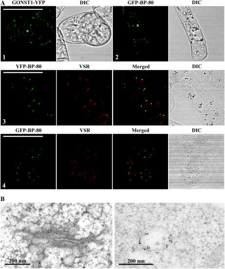
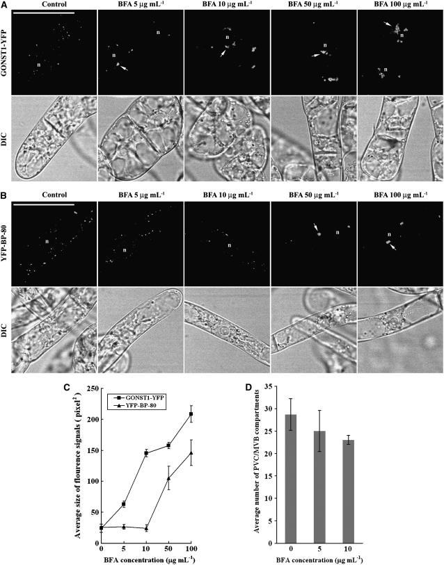
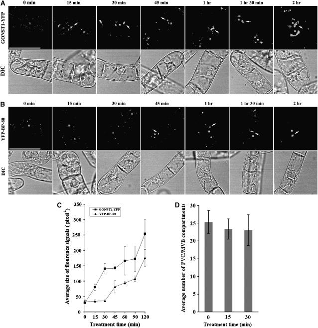
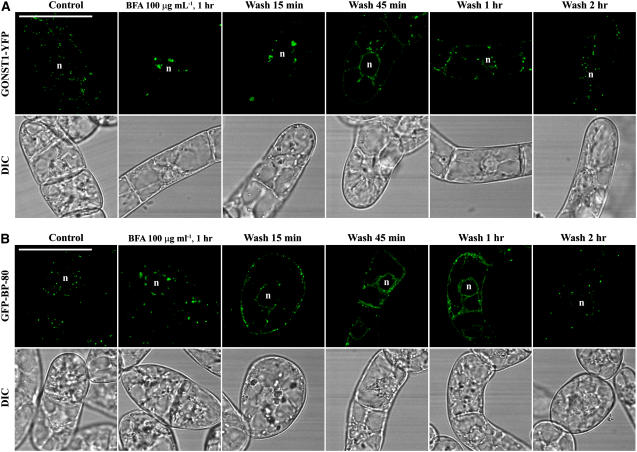
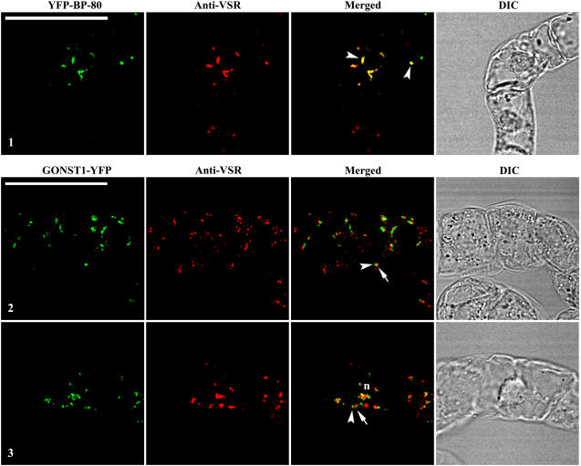
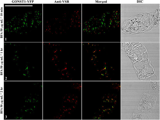
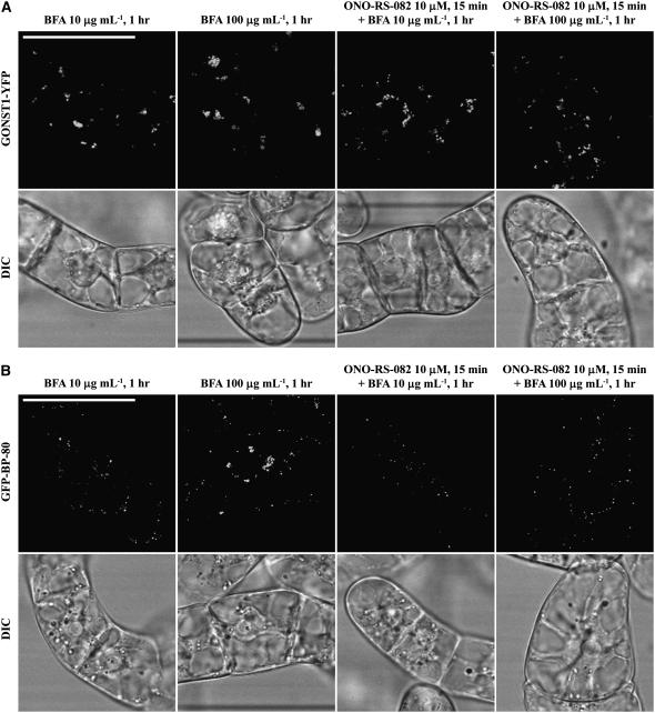
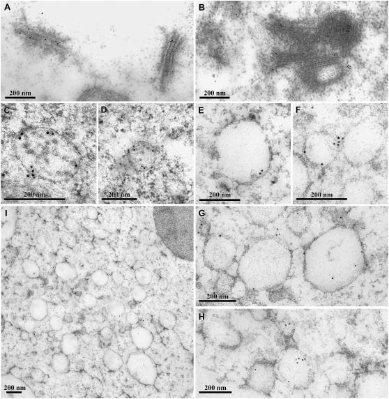
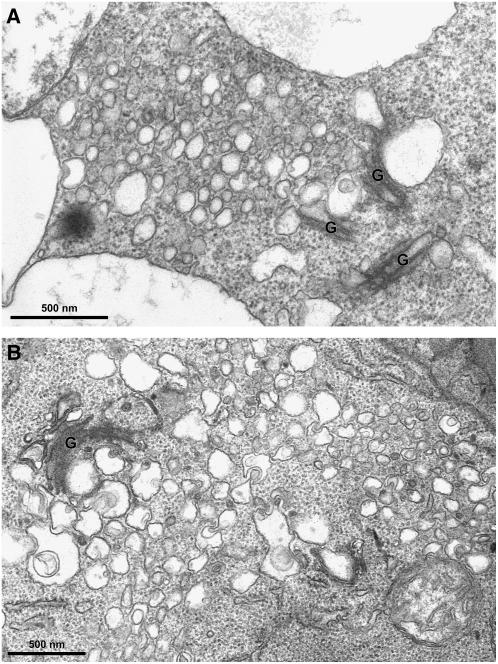
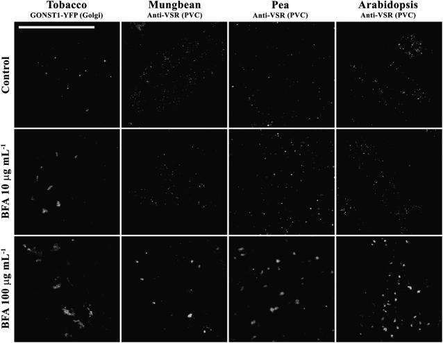
Little is known about the dynamics and molecular components of plant prevacuolar compartments (PVCs) in the secretory pathway. Using transgenic tobacco (Nicotiana tabacum) Bright-Yellow-2 (BY-2) cells expressing membrane-anchored yellow fluorescent protein (YFP) reporters marking Golgi or PVCs, we have recently demonstrated that PVCs are mobile multivesicular bodies defined by vacuolar sorting receptor proteins. Here, we demonstrate that Golgi and PVCs have different sensitivity in response to brefeldin A (BFA) treatment in living tobacco BY-2 cells. BFA at low concentrations (5-10 microg mL(-1)) induced YFP-marked Golgi stacks to form both endoplasmic reticulum-Golgi hybrid structures and BFA-induced aggregates, but had little effect on YFP-marked PVCs in transgenic BY-2 cells at both confocal and immunogold electron microscopy levels. However, BFA at high concentrations (50-100 microg mL(-1)) caused both YFP-marked Golgi stacks and PVCs to form aggregates in a dose- and time-dependent manner. Normal Golgi or PVC signals can be recovered upon removal of BFA from the culture media. Confocal immunofluorescence and immunogold electron microscopy studies with specific organelle markers further demonstrate that the PVC aggregates are distinct, but physically associated, with Golgi aggregates in BFA-treated cells and that PVCs might lose their internal vesicle structures at high BFA concentration. In addition, vacuolar sorting receptor-marked PVCs in root-tip cells of tobacco, pea (Pisum sativum), mung bean (Vigna radiata), and Arabidopsis (Arabidopsis thaliana) upon BFA treatment are also induced to form similar aggregates. Thus, we have demonstrated that the effects of BFA are not limited to endoplasmic reticulum and Golgi, but extend to PVC in the endomembrane system, which might provide a quick tool for distinguishing Golgi from PVC for its identification and characterization, as well as a possible new tool in studying PVC-mediated protein traffic in plant cells.


Figures

Figure 1.
Figure 1.
Subcellular localization of two reporters in transgenic BY-2 cells. A, Confocal immunofluorescent study of reporters. Similar punctate signals were detected from transgenic BY-2 cells expressing either the Golgi marker GONST1-YFP (image 1) or the PVC marker GFP-BP-80 (image 2), where both YFP BP-80 and GFP BP-80 reporter (green) largely colocalized with VSR antibodies (red). DIC images show the morphology of the tested cells. n, Nucleus. Bars = 50 μm. B, ImmunoEM localization of GONST1-YFP and GFP BP-80. Ultrathin sections prepared from high-pressure frozen/freeze-substituted transgenic GONST1-YFP BY-2 cells were labeled with either GFP antibodies (left) to detect the Golgi-localized GONST1-YFP reporter or VSR antibodies (right) to detect multivesicular PVCs. Bars = 200 nm.
Figure 2.
Figure 2.
BFA induces YFP-marked PVCs to form aggregates in a dose-dependent manner. A and B, Transgenic BY-2 cells expressing either the Golgi marker GONST1-YFP (A) or the PVC marker YFP BP-80 (B) were incubated with BFA at various concentrations, as indicated, for 1 h before they were collected for confocal imaging of YFP signals. Arrows indicate examples of BFA-induced aggregates in cells expressing these two reporters. DIC images show the morphology of the tested cells. n, Nucleus. Bars = 50 μm. C, Average sizes of the fluorescence signals of GONST1-YFP and YFP BP-80 in transgenic BY-2 cells treated with BFA at 50 μg mL−1 at indicated times (calculated using image J software on images of 500 × 500 pixels with 200 dpi). D, Average numbers of YFP BP-80-marked PVCs in transgenic BY-2 cells treated with BFA at indicated concentrations (0, 5, and 10 μg/mL) for 1 h.
Figure 3.
Figure 3.
Time-course formation of BFA-induced Golgi or PVC aggregates. A and B, Transgenic BY-2 cells expressing the Golgi marker GONST1-YFP (A) and the PVC marker YFP BP-80 (B) were first incubated with BFA at 50 μg mL−1, followed by sample collection at indicated times for confocal imaging of YFP signals. Arrows indicate examples of BFA-induced aggregates derived from either the YFP-marked Golgi (A) or the YFP-marked PVCs (B). DIC images show the morphology of the tested cells. n, Nucleus. Bars = 50 μm. C, Average sizes of the fluorescence signals of GONST1-YFP and YFP BP-80 in transgenic BY-2 cells treated with BFA at 50 μg mL−1 at indicated times (calculated using imageJ software on images of 500 × 500 pixels with 200 dpi). D, Average numbers of YFP BP-80-marked PVCs in a single transgenic BY-2 cell at indicated times upon BFA treatment at 50 μg/mL.
Figure 4.
Figure 4.
Recovery of BFA-induced aggregates. Transgenic BY-2 cells expressing the Golgi marker GONST1-YFP (A) and the PVC marker GFP BP-80 (B) were treated with BFA at 100 μg mL−1 for 1 h before BFA was washed off with fresh Murashige and Skoog medium, followed by sample collection at indicated times for confocal imaging. DIC images show the morphology of the tested cells. n, Nucleus. Bars = 50 μm.
Figure 5.
Figure 5.
BFA-induced PVC aggregates are distinct from Golgi aggregates. Transgenic BY-2 cells expressing the Golgi marker GONST1-YFP and the PVC marker YFP BP-80 were treated with BFA at 50 μg mL−1 for 1 h (images 1 and 2) or 2 h (image 3) prior to fixation. The fixed cells were then labeled with VSR antibodies to detect PVCs (red), whereas the YFP reporters (green) were ready for detection. Arrowheads in merged image 1 indicate examples of colocalization between the PVC reporter YFP BP-80 and VSR antibodies. Arrowheads and arrows in images 2 and 3 indicate examples of distinct, but closely associated, BFA-induced aggregates derived from Golgi (green) and PVC (red; termed Golgi-PVC hybrid). DIC images show the morphology of the tested cells. Bars = 50 μm.
Figure 6.
Figure 6.
Formation of Golgi-PVC hybrids was time dependent. Transgenic BY-2 cells expressing the Golgi reporter GONST1-YFP were treated with BFA at 50 μg mL−1, followed by sample collection at indicated time points and used for fixation. The fixed cells were then labeled with VSR antibodies to detect PVCs (red), whereas the YFP-marked Golgi (green) was ready for detection. DIC images show the morphology of the tested cells. Bar = 50 μm.
Figure 7.
Figure 7.
The target of BFA effect on Golgi and PVC is distinct. Transgenic BY-2 cells expressing the Golgi reporter GONST1-YFP (A) and PVC reporter GFP BP-80 (B) were treated with ONO-RS-082 at 10 μm for 15 min, followed by BFA treatment at 10 or 100 μg mL−1 for 1 h as indicated (images 3 and 4) before confocal imaging. Controls included BFA treatment at 10 or 100 μg mL−1 for 1 h before confocal imaging (images 1 and 2). DIC images show the morphology of the tested cells. Bars = 50 μm.
Figure 8.
Figure 8.
ImmunoEM identification of BFA-induced aggregates derived from Golgi and PVC. A, GFP antibodies labeled the Golgi stacks in untreated transgenic GONST1-YFP cells using ultrathin sections of Lowicryl (HM20). B, GFP antibodies labeled the curved Golgi structures in transgenic GONST1-YFP cells treated with BFA at 100 μg mL−1, using ultrathin sections of Lowicryl (HM20). C and D, Multivesicular PVCs as detected by VSR antibodies in untreated cells using ultrathin sections from high-pressure frozen/freeze-substituted material. E to H, PVC or PVC aggregates as detected by VSR antibodies in cells treated with BFA at 100 μg mL−1, using ultrathin samples of high-pressure frozen/freeze-substituted material. Bars = 200 nm. I, Overview of BFA-induced PVC aggregates in BY-2 cells treated with BFA at 100 μg mL−1 for 1 h, followed by high-pressure frozen/freeze substituted material. Bars = 500 nm.
Figure 9.
Figure 9.
Ultrastructural analysis of BFA-treated transgenic GONST1-YFP cells and Arabidopsis root-tip cells. A, Overview of BFA-induced Golgi and PVC aggregates in BY-2 cells treated with BFA at 100 μg mL−1 for 1 h. B, Overview of BFA-induced Golgi and PVC aggregates in Arabidopsis root tips treated with at BFA at 100 μg mL−1 for 1 h. Bars = 200 nm.
Figure 10.
Figure 10.
BFA also induced VSR-marked PVCs to form aggregates in other plant cell types. Root-tip cells from germinating seeds of transgenic GONST1-YFP tobacco, mung bean, pea, and Arabidopsis were treated with BFA at 0, 10, and 100 μg mL−1 for 1 h as indicated before they were either used for direct confocal imaging (for tobacco cells) to ready detect Golgi apparatus, or fixed and labeled with VSR antibodies (for pea, mung bean, and Arabidopsis root-tip cells) to detect PVCs. Bar = 50 μm.

Similar articles

Cited by

References

    1. Baldwin TC, Handford MG, Yuseff MI, Orellana A, Dupree P (2001) Identification and characterization of GONST1, a Golgi-localized GDP-mannose transporter in Arabidopsis. Plant Cell 13: 2283–2295 - PMC - PubMed
    1. Bassham DC, Gal S, daSilva CA, Raikhel NV (1995) An Arabidopsis syntaxin homologue isolated by functional complementation of a yeast pep12 mutant. Proc Natl Acad Sci USA 92: 7262–7266 - PMC - PubMed
    1. Bethke PC, Jones RL (2000) Vacuoles and prevacuolar compartments. Curr Opin Plant Biol 3: 469–475 - PubMed
    1. Boevink P, Oparka K, Santa CS, Martin B, Betteridge A, Hawes C (1998) Stacks on tracks: the plant Golgi apparatus traffics on an actin/ER network. Plant J 15: 441–447 - PubMed
    1. Bright NA, Lindsay MR, Stewart A, Luzio JP (2001) The relationship between lumenal and limiting membranes in swollen late endocytic compartments formed after wortmannin treatment or sucrose accumulation. Traffic 2: 631–642 - PubMed

Publication types

MeSH terms

LinkOut - more resources