. 2006 Dec;142(4):1427-41.
doi: 10.1104/pp.106.089169. Epub 2006 Oct 13.

Common plantain. A collection of expressed sequence tags from vascular tissue and a simple and efficient transformation method

Affiliations

Common plantain. A collection of expressed sequence tags from vascular tissue and a simple and efficient transformation method

Benjamin Pommerrenig et al. Plant Physiol. 2006 Dec.

Abstract

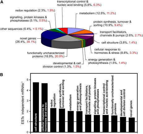
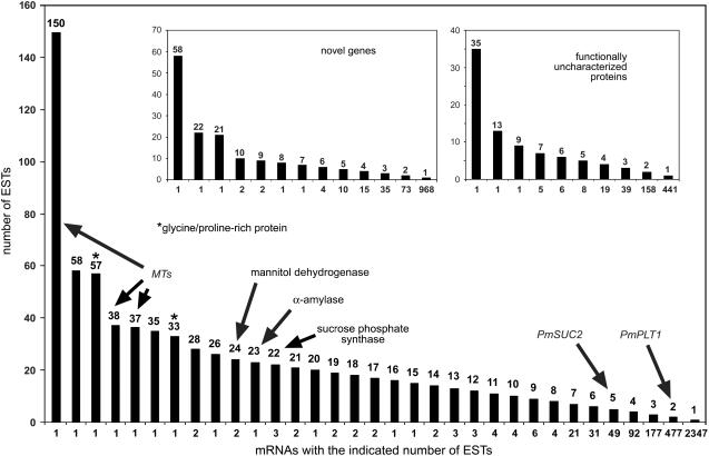
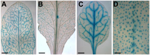
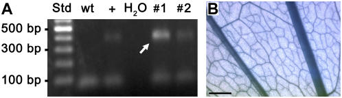
The vascular tissue of higher plants consists of specialized cells that differ from all other cells with respect to their shape and size, their organellar composition, their extracellular matrix, the type of their plasmodesmata, and their physiological functions. Intact and pure vascular tissue can be isolated easily and rapidly from leaf blades of common plantain (Plantago major), a plant that has been used repeatedly for molecular studies of phloem transport. Here, we present a transcriptome analysis based on 5,900 expressed sequence tags (ESTs) and 3,247 independent mRNAs from the Plantago vasculature. The vascular specificity of these ESTs was confirmed by the identification of well-known phloem or xylem marker genes. Moreover, reverse transcription-polymerase chain reaction, macroarray, and northern analyses revealed genes and metabolic pathways that had previously not been described to be vascular specific. Moreover, common plantain transformation was established and used to confirm the vascular specificity of a Plantago promoter-beta-glucuronidase construct in transgenic Plantago plants. Eventually, the applicability and usefulness of the obtained data were also demonstrated for other plant species. Reporter gene constructs generated with promoters from Arabidopsis (Arabidopsis thaliana) homologs of newly identified Plantago vascular ESTs revealed vascular specificity of these genes in Arabidopsis as well. The presented vascular ESTs and the newly developed transformation system represent an important tool for future studies of functional genomics in the common plantain vasculature.


Figures

Figure 1.
Figure 1.
Plantago vascular ESTs were divided into different groups. A, The obtained sequences were assigned either to one of 10 functional groups or to one of three groups standing for functionally uncharacterized proteins, for novel genes, or for a single mRNA with homology to sequences of viral origin (other sequences). The percentage of ESTs per group (black) and the percentage of independent mRNAs per group (= contigs plus singlets; red) are indicated. B, The EST-to-mRNA ratio was determined for 12 of the 13 groups shown in A. This ratio is a measure for the average strength of expression of the genes in a given group (higher bar = stronger average expression). The ratio is not shown for the other sequences group, which is represented by a single mRNA resulting from 26 ESTs.
Figure 2.
Figure 2.
Number of ESTs identified per individual mRNA. The number of identified and sequenced ESTs per mRNA is shown for all mRNAs (large image) and for the mRNAs of the novel genes and functionally uncharacterized proteins groups (insets). mRNAs for MTs, GRPs (asterisk), three biosynthetic enzymes (mannitol dehydrogenase, α-amylase, Suc phosphate synthase), or for previously characterized CC-specific transporters (PmSUC2, PmPLT1) are indicated.
Figure 3.
Figure 3.
Validation of vasculature-specific expression by northern blots and RT-PCR. A, Expression of four genes (PmMT1, PmMT2, PmRBCS1, and the contig P12.7.C1 [homologous to viral polyprotein]) in vascular (v) tissue isolated from leaves, in nonvascular (nv) tissue represented by petioles that had their vascular bundles extracted, and in intact petioles (p) was analyzed on northern blots. As expected, PmMT1 and PmMT2 are expressed almost exclusively in the vascular tissue, whereas expression of PmRBCS1 is higher in nonvascular tissue. Expression of P12.7.C1 is also highly specific for the vasculature. The bottom image represents the loading control (methylene blue-stained nylon filter). Gene names and accession numbers are given. B, Expression of 20 genes was analyzed in vascular (v) and nonvascular (nv) tissue. The figure shows the bands obtained by quantitative RT-PCR amplification with total RNA, the names of the respective genes (for novel genes the contig numbers are given), and their accession numbers. PmUBQ1 was used as a control that was expected to have similar expression levels in both tissues. PmPC1 (plastocyanine) was used as a control that was expected to be expressed mainly outside the vasculature. With the exception of PmQUA1, which shows comparable expression levels, all other genes are expressed mainly or exclusively in the vascular tissue.
Figure 4.
Figure 4.
Analysis of vascular tissue-specific expression using macroarrays. PCR-derived fragments of 216 different ESTs were spotted in duplicate on nylon filters and hybridized to radiolabeled mRNA isolated from vascular or nonvascular tissue. The relative expression intensities in vascular and nonvascular tissue are presented (±sd; n = 3). A, Genes with high expression levels. Bars marked with one asterisk represent mRNAs of MTs; the bar marked with two asterisks shows a vascular-specific aquaporin (PmAQP1); the bar marked with three asterisks represents a GRP (PmPSP1). B, Genes with medium expression levels. The bar marked with one asterisk represents PmAAMY1, an α-amylase gene; the bar marked with two asterisks shows PmSPS1, the gene of a Suc phosphate synthase. C, Genes with low expression levels. A to C, Thin arrows mark mRNAs from the functionally uncharacterized proteins group; thick and white arrows mark mRNAs from the novel genes group.
Figure 5.
Figure 5.
Analysis of the vascular specificity of Arabidopsis promoters chosen due to the vascular specificity of the homologous Plantago gene and of a Plantago promoter in Arabidopsis. A, GUS histochemical staining of the leaf of an AtMTK promoter-GUS plant. B, GUS histochemical staining of the leaf of an At2g16970 (tetracycline transporter-like protein) promoter-GUS plant. C, GUS histochemical staining of the leaf of a P12.0.C65 promoter-GUS plant. D, GUS histochemical staining of the leaf of an At5g62890 promoter-GUS plant. Scale bars = 2 mm in A, B, and D; 1 mm in C.
Figure 6.
Figure 6.
GUS histochemical staining of PmPLT1 promoter-GUS plants and identification of the GUS gene in genomic DNA of BASTA-resistant Plantago plants. A, PCR analysis of genomic DNA from two BASTA-resistant Plantago plants (nos. 1 and 2). The used primers amplify a 436-bp fragment of the GUS gene (arrow). The same fragment is seen only in a control with +. It is absent from wild-type (wt) Plantago and from the water (H2O) control. B, GUS histochemical staining of a leaf from a PmPLT1 promoter-GUS Plantago plant. Scale bar = 1 mm.

Similar articles

Cited by

References

    1. Aitchitt M, Ainsworth CC, Thangavelu M (1993) A rapid and efficient method for the extraction of total DNA from mature leaves of the date palm (Phoenix dactylifera L.). Plant Mol Biol Rep 11: 317–319
    1. Allona I, Quinn M, Shoop E, Swope K, St Cyr S, Carlis J, Riedl J, Retzel E, Campbell MM, Sederoff R, et al (1998) Analysis of xylem formation in pine by cDNA sequencing. Proc Natl Acad Sci USA 95: 9693–9698 - PMC - PubMed
    1. Altschul SF, Madden TL, Schaffer AA, Zhang J, Zhang Z, Miller W, Lipman DJ (1997) Gapped BLAST and PSI-BLAST: a new generation of protein database search programs. Nucleic Acids Res 25: 3389–3402 - PMC - PubMed
    1. Asano T, Masumura T, Kusano H, Kikuchi S, Kurita A, Shimada H, Kadowaki K (2002) Construction of a specialized cDNA library from plant cells isolated by laser capture microdissection: toward comprehensive analysis of the genes expressed in the rice phloem. Plant J 32: 401–408 - PubMed
    1. Balachandran S, Xiang Y, Schobert C, Thompson GA, Lucas WJ (1997) Phloem sap proteins from Cucurbita maxima and Ricinus communis have the capacity to traffic cell to cell through plasmodesmata. Proc Natl Acad Sci USA 94: 14150–14155 - PMC - PubMed

Publication types