Comparative Study
. 2006 Dec;142(4):1664-82.
doi: 10.1104/pp.106.087965. Epub 2006 Oct 13.

Heterologous expression and molecular and cellular characterization of CaPUB1 encoding a hot pepper U-Box E3 ubiquitin ligase homolog

Affiliations
Comparative Study

Heterologous expression and molecular and cellular characterization of CaPUB1 encoding a hot pepper U-Box E3 ubiquitin ligase homolog

Seok Keun Cho et al. Plant Physiol. 2006 Dec.

Abstract

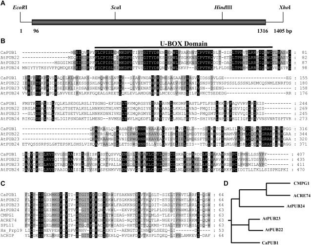
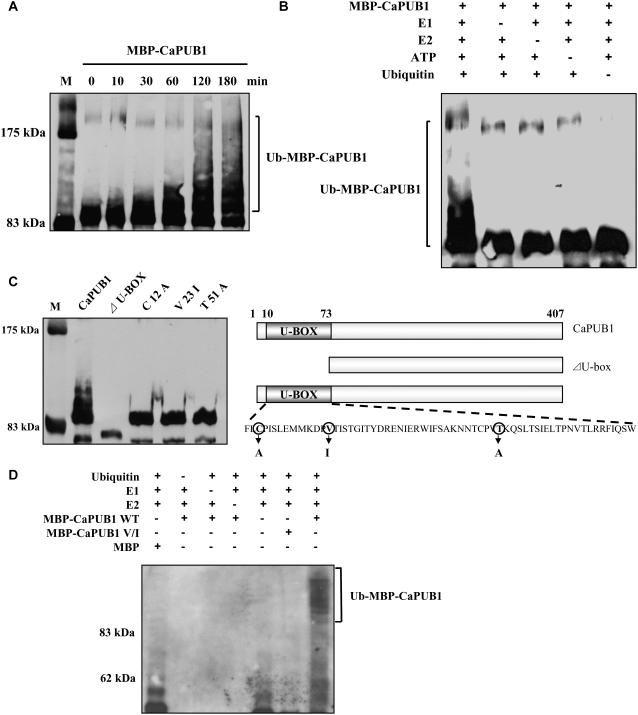
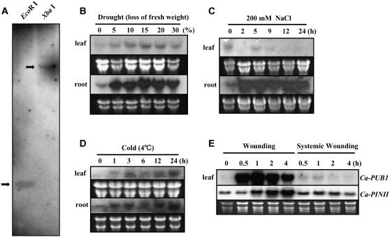
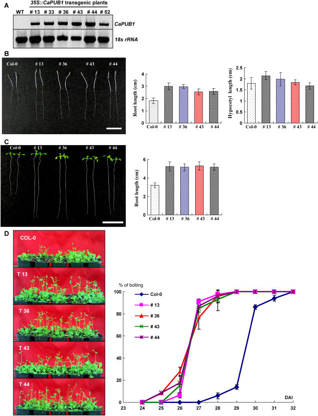
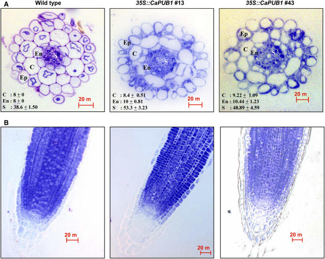
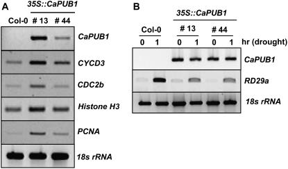
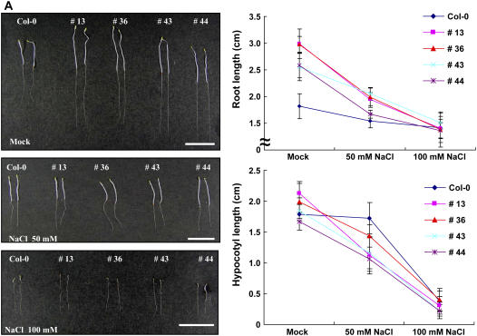
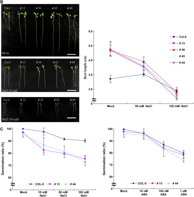
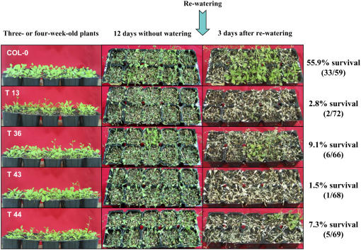
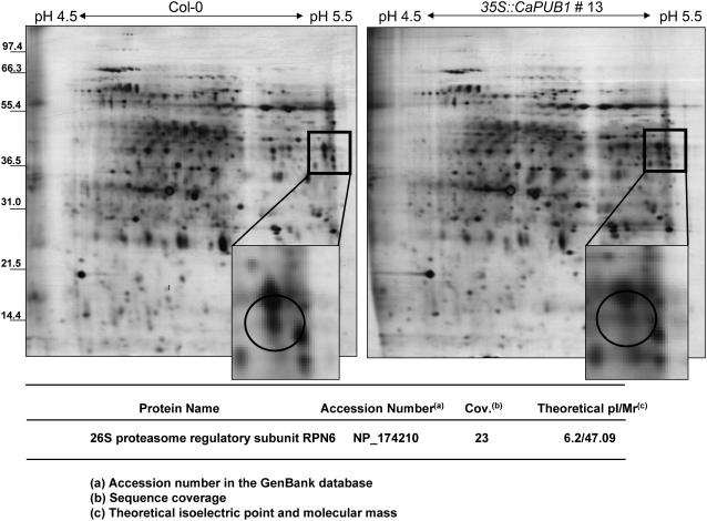
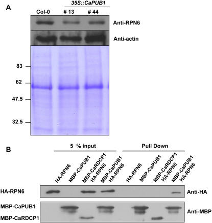
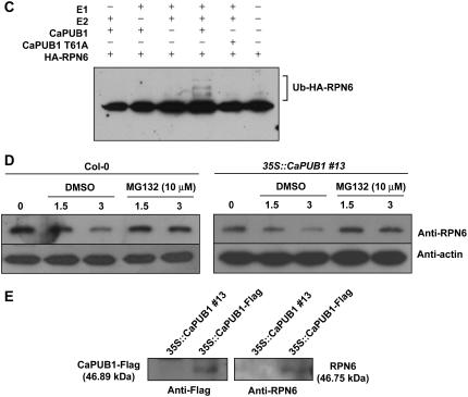
The U-box motif is a conserved domain found in the diverse isoforms of E3 ubiquitin ligase in eukaryotes. From water-stressed hot pepper (Capsicum annuum L. cv Pukang) plants, we isolated C. annuum putative U-box protein 1 (CaPUB1), which encodes a protein containing a single U-box motif in its N-terminal region. In vitro ubiquitination and site-directed mutagenesis assays revealed that CaPUB1 possessed E3 ubiquitin ligase activity and that the U-box motif was indeed essential for its enzyme activity. RNA gel-blot analysis showed that CaPUB1 mRNA was induced rapidly by a broad spectrum of abiotic stresses, including drought, high salinity, cold temperature, and mechanical wounding, but not in response to ethylene, abscisic acid, or a bacterial pathogen, suggesting its role in the early events in the abiotic-related defense response. Because transgenic work was extremely difficult in hot pepper, in this study we overexpressed CaPUB1 in Arabidopsis (Arabidopsis thaliana) to provide cellular information on the function of this gene in the development and plant responses to abiotic stresses. Transgenic Arabidopsis plants that constitutively expressed the CaPUB1 gene under the control of the cauliflower mosaic virus 35S promoter had markedly longer hypocotyls and roots and grew more rapidly than the wild type, leading to an early bolting phenotype. Microscopic analysis showed that 35S::CaPUB1 roots had increased numbers of small-sized cells, resulting in disordered, highly populated cell layers in the cortex, endodermis, and stele. In addition, CaPUB1-overexpressing plants displayed increased sensitivity to water stress and mild salinity. These results indicate that CaPUB1 is functional in Arabidopsis cells, thereby effectively altering cell and tissue growth and also the response to abiotic stresses. Comparative proteomic analysis showed that the level of RPN6 protein, a non-ATPase subunit of the 26S proteasome complex, was significantly reduced in 35SCaPUB1 seedlings as compared to the wild type. Pull-down and ubiquitination assays demonstrated that RPN6 interacted physically with CaPUB1 and was ubiquitinated in a CaPUB1-dependent manner in vitro. Although the physiological function of CaPUB1 is not yet clear, there are several possibilities for its involvement in a subset of physiological responses to counteract dehydration and high-salinity stresses in transgenic Arabidopsis seedlings.


Figures

Figure 1.
Figure 1.
Sequence analysis of hot pepper CaPUB1. A, Restriction enzyme map analysis of the hot pepper CaPUB1 cDNA clone. Solid bar depicts the coding region. Solid lines represent 5′- and 3′-untranslated regions. The sequence of pCaPUB1 has been deposited in the GenBank database under accession number DQ211901. B, Comparison of the derived amino acid sequence of hot pepper CaPUB1 with those of the Arabidopsis AtPUB22 (At3g52450), AtPUB23 (At2g35930), and AtPUB24 (At3g11840) U-box proteins. Amino acid residues that are conserved in at least three of the four sequences are shaded, whereas amino acids that are identical in all four proteins are shown in black. The solid line represents the U-box motif, which is essential for E3 Ub ligase activity. Dashes show gaps in the amino acid sequences that were introduced to optimize alignment. C, Sequence alignment of the U-box domain of CaPUB1 and other U-box proteins. The sequences of U-box motifs in hot pepper CaPUB1, Arabidopsis AtPUB22, AtPUB23, and AtPUB24, tobacco ACRE74 (GenBank accession no. AAP03884), parsley CMPG1 (Kirsch et al., 2001), rice SPL11 (Zeng et al., 2004), and human hCHIP and Prp19 (Ohi et al., 2003) are shown. Amino acid residues that are conserved in at least seven of the nine sequences are shaded. Amino acids that are identical in all nine proteins are shown in black. The numbers on the right indicate the amino acid residues. Dashes show gaps in the amino acid sequences that were introduced to optimize alignment. D, Phylogenetic relationship of U-box Ub-ligase homologs from hot pepper (CaPUB1), Arabidopsis (AtPUB22, AtPUB23, and AtPUB24), parsley (CMPG1), and tobacco (ACRE74).
Figure 2.
Figure 2.
In vitro ubiquitination assay of CaPUB1. A, The MBP-CaPUB1 fusion protein expressed in E. coli was incubated at 30°C for the indicated time points in the presence of E1, E2, ATP, and Ub. Samples were resolved by 8% SDS-PAGE and subjected to immunoblot analysis with the anti-MBP antibody. B, MBP-CaPUB1 was incubated at 30°C for 120 min in the presence or absence of E1, E2, ATP, and/or Ub. Samples were identically analyzed as described above. Ub ligase enzyme activity was not detected in the absence of any of the E1, E2, ATP, or Ub. C, The U-box motif is essential for E3 Ub ligase activity. Wild-type MBP-CaPUB1 and its various mutants were used in the Ub ligase enzyme assays. C12A, V23I, and T51A are mutants of MBP-CaPUB1 in which the conserved Cys-12, Val-23, and Thr-51 residues are replaced with Ala, Ile, and Ala, respectively. ΔU-BOX is a deletion mutant in which the N-terminal U-box domain is truncated. D, In vitro ubiquitination assay of the wild-type and V → I mutant MBP-CaPUB1 was repeated using an anti-Ub antibody.
Figure 3.
Figure 3.
Hybridization analysis of CaPUB1 genomic DNA and mRNA. A, Genomic Southern-blot analysis of the CaPUB1 gene. Hot pepper genomic DNA (10 μg/lane) was isolated from leaf tissue, digested with either EcoRI or XbaI and resolved on a 0.7% agarose gel. DNA on the gel was transferred to a nylon membrane filter. The filter was hybridized with 32P-labeled pCaPUB1 under high-stringency conditions. B to E, Light-grown 2-week-old hot pepper plants were subjected to drought (0%–30% loss of fresh weight; B), NaCl (200 mm; C), cold temperature (4°C; D), or mechanical wounding (E). Treated tissues were harvested at the indicated time points and total RNAs were isolated. Total RNAs (20 μg) were separated by electrophoresis on a 1% formaldehyde-agarose gel and blotted to a Hybond-N nylon membrane. To ensure equal loading of the RNA, the gel was stained with ethidium bromide after electrophoresis. To confirm complete transfer of RNA to the membrane filter, both gel and membrane were viewed under UV light after transfer. The filter was hybridized with 32P-labeled pCaPUB1or pCaPINII under high-stringency conditions.
Figure 4.
Figure 4.
Molecular characterization and phenotype of CaPUB1-overexpressing transgenic Arabidopsis plants. A, RT-PCR analysis of wild-type and 35SCaPUB1 transgenic lines. B and C, Morphological comparisons of the wild-type and CaPUB1-overexpressing lines under dark (B) or light (C) growth conditions. Bar = 15 mm. D, Early bolting phenotype of 35SCaPUB1 relative to the wild-type plant. [See online article for color version of this figure.]
Figure 5.
Figure 5.
Cellular phenotypes of wild-type and 35SCaPUB1 roots. A, Transverse sections of roots from 5-d-old light-grown wild-type and 35SCaPUB1 seedlings (lines 13 and 43) stained with toluidine. Average number of cells in the cortex, endodermis, and stele is indicated. Ep, Epidermis; C, cortex; En, endodermis; S, stele. Bar = 20 μm. B, Longitudinal sections of roots from 5-d-old light-grown wild-type and 35SCaPUB1 seedlings (lines 13 and 43) stained with toluidine. Bar = 20 μm. [See online article for color version of this figure.]
Figure 6.
Figure 6.
Expression of cell cycle-related and drought-induced genes in the 35SCaPUB1 plants. A, Transcription of cell-cycle-regulated genes was determined by RT-PCR. Wild-type and two independent 35SCaPUB1 transgenic lines (13 and 44) were analyzed. The cell cycle-related genes are the D-type cyclin CycD3, cell cycle-dependent kinase-related gene CDC2b, S-phase-specific PCNA, and chromosome assembly histone H3. As a negative control, the 18S rRNA transcript level was shown. B, Induction level of the RD29a gene in the wild type and two independent 35SCaPUB1 transgenic lines (13 and 44) in response to drought stress. Light-grown 10-d-old wild-type and 35SCaPUB1 transgenic seedlings were dehydrated for 1 h on Whatman 3MM filter paper at room temperature. Induction level of RD29a, a typical drought stress-induced gene (Liu et al., 1998), was examined by RT-PCR. The 18S rRNA transcript level was used as a loading control.
Figure 7.
Figure 7.
Increased sensitivity of 35SCaPUB1 transgenic lines to salt stress. A, Five-day-old etiolated wild-type and transgenic seedlings were incubated with 50 to 100 mm NaCl and root and hypocotyl growth assays were carried out. The values are means ± sd (n = 4). Bar = 15 mm. B, Seven-day-old light-grown wild-type and transgenic seedlings were subjected to 50 to 100 mm NaCl and the growth patterns of roots and hypocotyls were monitored. The values are means ± sd (n = 4). Bar = 15 mm. C, Germination ratio of wild-type and 35SCaPUB1 transgenic lines in the absence or presence of NaCl (10–100 mm; left); germination ratio of wild-type and 35SCaPUB1 transgenic lines in the absence or presence of ABA (0.01–1 μm; right). [See online article for color version of this figure.]
Figure 7.
Figure 7.
Increased sensitivity of 35SCaPUB1 transgenic lines to salt stress. A, Five-day-old etiolated wild-type and transgenic seedlings were incubated with 50 to 100 mm NaCl and root and hypocotyl growth assays were carried out. The values are means ± sd (n = 4). Bar = 15 mm. B, Seven-day-old light-grown wild-type and transgenic seedlings were subjected to 50 to 100 mm NaCl and the growth patterns of roots and hypocotyls were monitored. The values are means ± sd (n = 4). Bar = 15 mm. C, Germination ratio of wild-type and 35SCaPUB1 transgenic lines in the absence or presence of NaCl (10–100 mm; left); germination ratio of wild-type and 35SCaPUB1 transgenic lines in the absence or presence of ABA (0.01–1 μm; right). [See online article for color version of this figure.]
Figure 8.
Figure 8.
Increased sensitivity of 35SCaPUB1 transgenic lines to water stress. Wild-type and transgenic Arabidopsis plants were grown in pots for 4 weeks. Water was withheld for 12 d, followed by rewatering for 3 d. Dehydration sensitivity was assayed as the capability of plants to resume growth when returned to normal conditions following water stress. [See online article for color version of this figure.]
Figure 9.
Figure 9.
Silver-stained 2-DE map of wild-type and 35SCaPUB1 transgenic Arabidopsis seedlings. Protein samples were prepared from 7-d-old whole seedlings as described in “Materials and Methods.” Proteins (350 μg) were applied to an immobilized pH gradient strip by the in-gel rehydration method. Subsequently, 9% to 16% gradient SDS-PAGE was performed to separate proteins in the second dimension. The proteins were visualized with silver nitrate. The arrow in the magnified region of the 2-DE image shows a protein spot whose abundance is clearly reduced in the 35SCaPUB1 transgenic line as compared to the wild type. This spot was excised from the gel and analyzed by MALDI-TOF MS.
Figure 10.
Figure 10.
CaPUB1 interacts with the RPN6 protein. A, Immunoblot analysis of RPN6 protein. Protein samples (20 μg of total proteins) were prepared from 7-d-old wild-type and two independent transgenic seedlings (lines 13 and 44), and subjected to immunoblot analysis using an anti-RPN6 antibody or anti-actin antibody as a negative control. The blot was visualized by the chemiluminescence detection method. The equivalence of protein loading among lanes of the SDS-PAGE gel was demonstrated by Coomassie Brilliant Blue R-250 staining of the proteins on the gel. B, In vitro pull-down assay. MBP-CaPUB1 and HA-RPN6 fusion proteins were coincubated with amylose affinity matrix. The bound protein was eluted from the amylose resin by 10 mm maltose, resolved by 12.5% SDS-PAGE, and transferred to a nitrocellulose membrane. The blot was probed with an anti-HA antibody or anti-MBP antibody. C, In vitro ubiquitination assay of RPN6 using CaPUB1 as the E3 Ub ligase. Recombinant HA-RPN6 protein was incubated in the presence or absence of Ub, ATP, E1, E2, and wild-type MBP-CaPUB1 or its mutant form (T51A), for the appropriate time periods, and subjected to immunoblotting using an anti-HA antibody. D, In vivo interaction of CaPUB1 and RPN6. The wild-type (left) and 35SCaPUB1 transgenic seedlings (right) were treated with 100 μm cycloheximide for different time periods (0–3 h) in the absence or presence of MG132, an inhibitor of the 26S proteasome. During incubation, changes in the level of RPN6 protein were monitored using an anti-RPN6 antibody. E, In vivo immunoprecipitation assay. Putative interacting proteins with CaPUB1 were immunoprecipitated from the 35SCaPUB1-Flag transgenic seedlings using an anti-Flag antibody. Immunoprecipitated proteins were subsequently analyzed by immunoblotting with an anti-RPN antibody. As a specificity control, 35SCaPUB1 seedlings were used in an identical experiment. [See online article for color version of this figure.]
Figure 10.
Figure 10.
CaPUB1 interacts with the RPN6 protein. A, Immunoblot analysis of RPN6 protein. Protein samples (20 μg of total proteins) were prepared from 7-d-old wild-type and two independent transgenic seedlings (lines 13 and 44), and subjected to immunoblot analysis using an anti-RPN6 antibody or anti-actin antibody as a negative control. The blot was visualized by the chemiluminescence detection method. The equivalence of protein loading among lanes of the SDS-PAGE gel was demonstrated by Coomassie Brilliant Blue R-250 staining of the proteins on the gel. B, In vitro pull-down assay. MBP-CaPUB1 and HA-RPN6 fusion proteins were coincubated with amylose affinity matrix. The bound protein was eluted from the amylose resin by 10 mm maltose, resolved by 12.5% SDS-PAGE, and transferred to a nitrocellulose membrane. The blot was probed with an anti-HA antibody or anti-MBP antibody. C, In vitro ubiquitination assay of RPN6 using CaPUB1 as the E3 Ub ligase. Recombinant HA-RPN6 protein was incubated in the presence or absence of Ub, ATP, E1, E2, and wild-type MBP-CaPUB1 or its mutant form (T51A), for the appropriate time periods, and subjected to immunoblotting using an anti-HA antibody. D, In vivo interaction of CaPUB1 and RPN6. The wild-type (left) and 35SCaPUB1 transgenic seedlings (right) were treated with 100 μm cycloheximide for different time periods (0–3 h) in the absence or presence of MG132, an inhibitor of the 26S proteasome. During incubation, changes in the level of RPN6 protein were monitored using an anti-RPN6 antibody. E, In vivo immunoprecipitation assay. Putative interacting proteins with CaPUB1 were immunoprecipitated from the 35SCaPUB1-Flag transgenic seedlings using an anti-Flag antibody. Immunoprecipitated proteins were subsequently analyzed by immunoblotting with an anti-RPN antibody. As a specificity control, 35SCaPUB1 seedlings were used in an identical experiment. [See online article for color version of this figure.]

Similar articles

Cited by

References

    1. Azevedo C, Santos-Rosa MJ, Shirasu K (2001) The U-box protein family in plants. Trends Plant Sci 6: 354–358 - PubMed
    1. Baumeister W, Walz J, Zuhl F, Seemuller E (1998) The proteasome: paradigm of a self-compartmentalizing protease. Cell 92: 367–380 - PubMed
    1. Boyer JS (1982) Plant productivity and environment. Science 218: 443–448 - PubMed
    1. Bray EA (1997) Plant responses to water deficit. Trends Plant Sci 2: 48–54
    1. Cho SK, Kim JE, Park J-A, Eom TJ, Kim WT (2006) Constitutive expression of abiotic stress-inducible hot pepper CaXTH3, which encodes a xyloglucan endotransglucosylase/hydrolase homolog, improves drought and salt tolerance in transgenic Arabidopsis plants. FEBS Lett 580: 3136–3144 - PubMed

Publication types

MeSH terms

Associated data