

Characterization of the Moraxella catarrhalis opa-like protein, OlpA, reveals a phylogenetically conserved family of outer membrane proteins
- PMID: 17041038
- PMCID: PMC1797210
- DOI: 10.1128/JB.00788-06
Characterization of the Moraxella catarrhalis opa-like protein, OlpA, reveals a phylogenetically conserved family of outer membrane proteins
Abstract
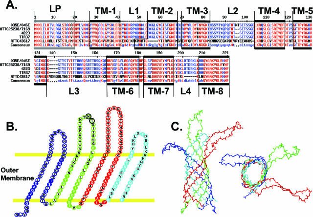
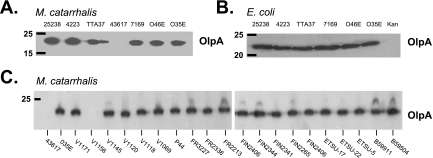
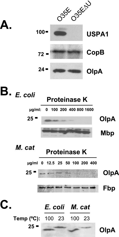
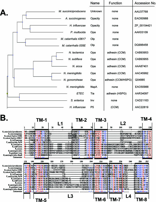
Moraxella catarrhalis is a human-restricted pathogen that can cause respiratory tract infections. In this study, we identified a previously uncharacterized 24-kDa outer membrane protein with a high degree of similarity to Neisseria Opa protein adhesins, with a predicted beta-barrel structure consisting of eight antiparallel beta-sheets with four surface-exposed loops. In striking contrast to the antigenically variable Opa proteins, the M. catarrhalis Opa-like protein (OlpA) is highly conserved and constitutively expressed, with 25 of 27 strains corresponding to a single variant. Protease treatment of intact bacteria and isolation of outer membrane vesicles confirm that the protein is surface exposed yet does not bind host cellular receptors recognized by neisserial Opa proteins. Genome-based analyses indicate that OlpA and Opa derive from a conserved family of proteins shared by a broad array of gram-negative bacteria.
Figures
Similar articles
-
Characterization of a novel porin protein from Moraxella catarrhalis and identification of an immunodominant surface loop.J Bacteriol. 2005 Sep;187(18):6528-35. doi: 10.1128/JB.187.18.6528-6535.2005. J Bacteriol. 2005. PMID: 16159786 Free PMC article.
-
Identification and characterization of a novel outer membrane protein (OMP J) of Moraxella catarrhalis that exists in two major forms.J Bacteriol. 2005 Dec;187(23):7977-84. doi: 10.1128/JB.187.23.7977-7984.2005. J Bacteriol. 2005. PMID: 16291671 Free PMC article.
-
Identification and characterization of outer membrane proteins G1a and G1b of Moraxella catarrhalis.Vaccine. 2004 Jun 30;22(20):2533-40. doi: 10.1016/j.vaccine.2003.12.016. Vaccine. 2004. PMID: 15193378
-
Moraxella (Branhamella) catarrhalis--clinical and molecular aspects of a rediscovered pathogen.J Med Microbiol. 1997 May;46(5):360-71. doi: 10.1099/00222615-46-5-360. J Med Microbiol. 1997. PMID: 9152030 Review.
-
MID and UspA1/A2 of the human respiratory pathogen Moraxella catarrhalis, and interactions with the human host as basis for vaccine development.Acta Biochim Pol. 2006;53(3):445-56. Epub 2006 Sep 9. Acta Biochim Pol. 2006. PMID: 16964325 Review.
Cited by
-
Panel 5: Microbiology and immunology panel.Otolaryngol Head Neck Surg. 2013 Apr;148(4 Suppl):E64-89. doi: 10.1177/0194599812459636. Otolaryngol Head Neck Surg. 2013. PMID: 23536533 Free PMC article. Review.
-
Vaccine targets against Moraxella catarrhalis.Expert Opin Ther Targets. 2016;20(1):19-33. doi: 10.1517/14728222.2015.1081686. Epub 2015 Aug 26. Expert Opin Ther Targets. 2016. PMID: 26565427 Free PMC article. Review.
-
Potential impact of a Moraxella catarrhalis vaccine in COPD.Vaccine. 2019 Sep 3;37(37):5551-5558. doi: 10.1016/j.vaccine.2016.12.066. Epub 2017 Feb 6. Vaccine. 2019. PMID: 28185742 Free PMC article. Review.
-
A Moraxella catarrhalis vaccine to protect against otitis media and exacerbations of COPD: An update on current progress and challenges.Hum Vaccin Immunother. 2017 Oct 3;13(10):2322-2331. doi: 10.1080/21645515.2017.1356951. Hum Vaccin Immunother. 2017. PMID: 28853985 Free PMC article. Review.
-
The modA10 phasevarion of nontypeable Haemophilus influenzae R2866 regulates multiple virulence-associated traits.Microb Pathog. 2016 Mar;92:60-67. doi: 10.1016/j.micpath.2015.12.006. Epub 2015 Dec 21. Microb Pathog. 2016. PMID: 26718097 Free PMC article.
References
-
- Bandak, S. I., M. R. Turnak, B. S. Allen, L. D. Bolzon, D. A. Preston, S. K. Bouchillon, and D. J. Hoban. 2001. Antibiotic susceptibilities among recent clinical isolates of Haemophilus influenzae and Moraxella catarrhalis from fifteen countries. Eur. J. Clin. Microbiol. Infect. Dis. 20:55-60. - PubMed
-
- Bates, P. A., L. A. Kelley, R. M. MacCallum, and M. J. Sternberg. 2001. Enhancement of protein modeling by human intervention in applying the automatic programs 3D-JIGSAW and 3D-PSSM. Proteins 5(Suppl.):39-46. - PubMed
-
- Bates, P. A., and M. J. Sternberg. 1999. Model building by comparison at CASP3: using expert knowledge and computer automation. Proteins 3(Suppl.):47-54. - PubMed
-
- Christensen, J. J. 1999. Moraxella (Branhamella) catarrhalis: clinical, microbiological and immunological features in lower respiratory tract infections. APMIS 88(Suppl.):1-36. - PubMed
Publication types
MeSH terms
Substances
Grants and funding
LinkOut - more resources
Full Text Sources
Other Literature Sources
Molecular Biology Databases
