

Complete sequence analysis of novel plasmids from emetic and periodontal Bacillus cereus isolates reveals a common evolutionary history among the B. cereus-group plasmids, including Bacillus anthracis pXO1
- PMID: 17041058
- PMCID: PMC1797222
- DOI: 10.1128/JB.01313-06
Complete sequence analysis of novel plasmids from emetic and periodontal Bacillus cereus isolates reveals a common evolutionary history among the B. cereus-group plasmids, including Bacillus anthracis pXO1
Abstract
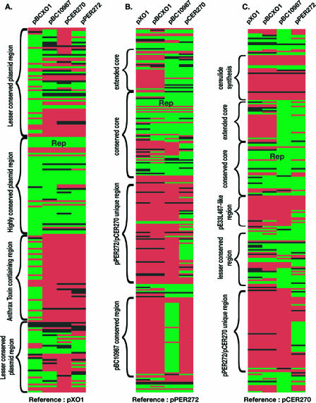
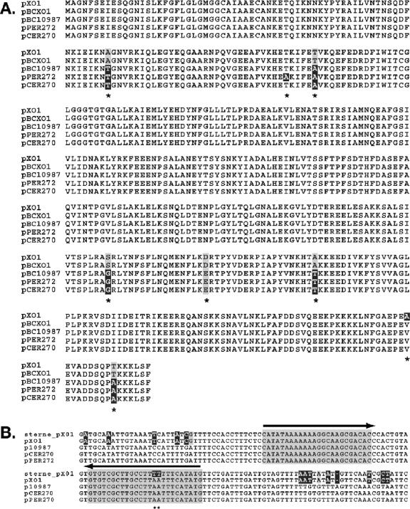
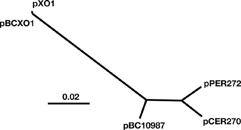
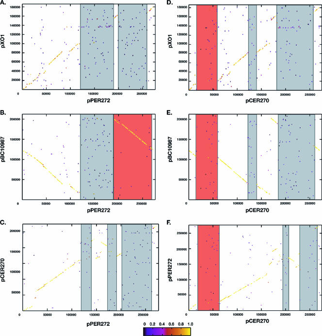
The plasmids of the members of the Bacillus cereus sensu lato group of organisms are essential in defining the phenotypic traits associated with pathogenesis and ecology. For example, Bacillus anthracis contains two plasmids, pXO1 and pXO2, encoding toxin production and encapsulation, respectively, that define this species pathogenic potential, whereas the presence of a Bt toxin-encoding plasmid defines Bacillus thuringiensis isolates. In this study the plasmids from B. cereus isolates that produce emetic toxin or are linked to periodontal disease were sequenced and analyzed. Two periodontal isolates examined contained almost identical approximately 272-kb plasmids, named pPER272. The emetic toxin-producing isolate contained one approximately 270-kb plasmid, named pCER270, encoding the cereulide biosynthesis gene cluster. Comparative sequence analyses of these B. cereus plasmids revealed a high degree of sequence similarity to the B. anthracis pXO1 plasmid, especially in a putative replication region. These plasmids form a newly defined group of pXO1-like plasmids. However, these novel plasmids do not contain the pXO1 pathogenicity island, which in each instance is replaced by plasmid specific DNA. Plasmids pCER270 and pPER272 share regions that are not found in any other pXO1-like plasmids. Evolutionary studies suggest that these plasmids are more closely related to each other than to other identified B. cereus plasmids. Screening of a population of B. cereus group isolates revealed that pXO1-like plasmids are more often found in association with clinical isolates. This study demonstrates that the pXO1-like plasmids may define pathogenic B. cereus isolates in the same way that pXO1 and pXO2 define the B. anthracis species.
Figures
Similar articles
-
Cereulide synthetase gene cluster from emetic Bacillus cereus: structure and location on a mega virulence plasmid related to Bacillus anthracis toxin plasmid pXO1.BMC Microbiol. 2006 Mar 2;6:20. doi: 10.1186/1471-2180-6-20. BMC Microbiol. 2006. PMID: 16512902 Free PMC article.
-
Differentiation and characterization by molecular techniques of Bacillus cereus group isolates from poto poto and dégué, two traditional cereal-based fermented foods of Burkina Faso and Republic of Congo.J Food Prot. 2007 May;70(5):1165-73. doi: 10.4315/0362-028x-70.5.1165. J Food Prot. 2007. PMID: 17536675
-
Distribution, diversity, and potential mobility of extrachromosomal elements related to the Bacillus anthracis pXO1 and pXO2 virulence plasmids.Appl Environ Microbiol. 2009 May;75(10):3016-28. doi: 10.1128/AEM.02709-08. Epub 2009 Mar 20. Appl Environ Microbiol. 2009. PMID: 19304837 Free PMC article.
-
What sets Bacillus anthracis apart from other Bacillus species?Annu Rev Microbiol. 2009;63:451-76. doi: 10.1146/annurev.micro.091208.073255. Annu Rev Microbiol. 2009. PMID: 19514852 Review.
-
Bacillus anthracis genetics and virulence gene regulation.Curr Top Microbiol Immunol. 2002;271:143-64. doi: 10.1007/978-3-662-05767-4_7. Curr Top Microbiol Immunol. 2002. PMID: 12224521 Review.
Cited by
-
Bacillus cereus Biofilms-Same, Only Different.Front Microbiol. 2016 Jul 7;7:1054. doi: 10.3389/fmicb.2016.01054. eCollection 2016. Front Microbiol. 2016. PMID: 27458448 Free PMC article. Review.
-
Comparative Genomics of Marine Sponge-Derived Streptomyces spp. Isolates SM17 and SM18 With Their Closest Terrestrial Relatives Provides Novel Insights Into Environmental Niche Adaptations and Secondary Metabolite Biosynthesis Potential.Front Microbiol. 2019 Jul 26;10:1713. doi: 10.3389/fmicb.2019.01713. eCollection 2019. Front Microbiol. 2019. PMID: 31404169 Free PMC article.
-
Identification of the main promoter directing cereulide biosynthesis in emetic Bacillus cereus and its application for real-time monitoring of ces gene expression in foods.Appl Environ Microbiol. 2010 Feb;76(4):1232-40. doi: 10.1128/AEM.02317-09. Epub 2009 Dec 28. Appl Environ Microbiol. 2010. PMID: 20038713 Free PMC article.
-
Insights into the environmental resistance gene pool from the genome sequence of the multidrug-resistant environmental isolate Escherichia coli SMS-3-5.J Bacteriol. 2008 Oct;190(20):6779-94. doi: 10.1128/JB.00661-08. Epub 2008 Aug 15. J Bacteriol. 2008. PMID: 18708504 Free PMC article.
-
Bacillus anthracis physiology and genetics.Mol Aspects Med. 2009 Dec;30(6):386-96. doi: 10.1016/j.mam.2009.07.004. Epub 2009 Aug 3. Mol Aspects Med. 2009. PMID: 19654018 Free PMC article. Review.
References
-
- Agata, N., M. Ohta, and M. Mori. 1996. Production of an emetic toxin, cereulide, is associated with a specific class of Bacillus cereus. Curr. Microbiol. 33:67-69. - PubMed
-
- Agata, N., M. Ohta, M. Mori, and M. Isobe. 1995. A novel dodecadepsipeptide, cereulide, is an emetic toxin of Bacillus cereus. FEMS Microbiol. Lett. 129:17-19. - PubMed
-
- Apetroaie, C., M. A. Andersson, C. Sproer, I. Tsitko, R. Shaheen, E. L. Jaaskelainen, L. M. Wijnands, R. Heikkila, and M. S. Salkinoja-Salonen. 2005. Cereulide-producing strains of Bacillus cereus show diversity. Arch. Microbiol. 184:141-151. - PubMed
-
- Barker, M., B. Thakker, and F. G. Priest. 2005. Multilocus sequence typing reveals that Bacillus cereus strains isolated from clinical infections have distinct phylogenetic origins. FEMS Microbiol. Lett. 245:179-184. - PubMed
Publication types
MeSH terms
Substances
Grants and funding
LinkOut - more resources
Full Text Sources
Other Literature Sources
