. 2006 Oct;18(10):2650-63.
doi: 10.1105/tpc.106.046110. Epub 2006 Oct 13.

A pentatricopeptide repeat protein facilitates the trans-splicing of the maize chloroplast rps12 pre-mRNA

Affiliations

A pentatricopeptide repeat protein facilitates the trans-splicing of the maize chloroplast rps12 pre-mRNA

Christian Schmitz-Linneweber et al. Plant Cell. 2006 Oct.

Abstract

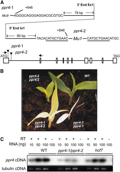
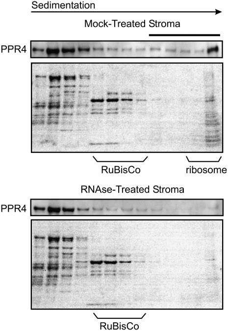
The pentatricopeptide repeat (PPR) is a degenerate 35-amino acid repeat motif that is widely distributed among eukaryotes. Genetic, biochemical, and bioinformatic data suggest that many PPR proteins influence specific posttranscriptional steps in mitochondrial or chloroplast gene expression and that they may typically bind RNA. However, biological functions have been determined for only a few PPR proteins, and with few exceptions, substrate RNAs are unknown. To gain insight into the functions and substrates of the PPR protein family, we characterized the maize (Zea mays) nuclear gene ppr4, which encodes a chloroplast-targeted protein harboring both a PPR tract and an RNA recognition motif. Microarray analysis of RNA that coimmunoprecipitates with PPR4 showed that PPR4 is associated in vivo with the first intron of the plastid rps12 pre-mRNA, a group II intron that is transcribed in segments and spliced in trans. ppr4 mutants were recovered through a reverse-genetic screen and shown to be defective for rps12 trans-splicing. The observations that PPR4 is associated in vivo with rps12-intron 1 and that it is also required for its splicing demonstrate that PPR4 is an rps12 trans-splicing factor. These findings add trans-splicing to the list of RNA-related functions associated with PPR proteins and suggest that plastid group II trans-splicing is performed by different machineries in vascular plants and algae.


Figures

Figure 1.
Figure 1.
Alignment of PPR4 Orthologs in Maize (Zm), Rice (Os), and Arabidopsis (At). TargetP (Emanuelsson et al., 2000) and Predotar (Small et al., 2004) algorithms both predict that all three proteins are targeted to the chloroplast. The insertion site of the Mu9 element in ppr4-1 mutants is indicated by an arrowhead. The RRM domain is underlined, with the thicker lines denoting its two core RNP motifs. PPR repeats are indicated below the sequences by double-headed arrows. At PPR4 corresponds to Arabidopsis Genome Initiative locus At5g04810; Os PPR4 corresponds to The Institute for Genomic Research locus Os04g58780.
Figure 2.
Figure 2.
Mu-Induced ppr4 Mutants. (A) Positions of the Mu insertions in ppr4-1 and ppr4-2. The DNA sequence flanking the Mu insertions is shown, with the intron-exon border indicated by a dotted line. Residue numbers with respect to the start codon in the genomic sequence are indicated. A 10-nucleotide duplication flanking the ppr4-2 insertion site is underlined; this sequence is present only once in the progenitor allele. The type of Mu element at each site (i.e., Mu1 or Mu9) was deduced from polymorphisms in the terminal inverted repeat sequences. A schematic representation of the ppr4 gene is shown below. Boxes represent exons, and lines represent introns (drawn to scale). Mu element insertion sites are marked. Arrows illustrate the primers used for RT-PCR analysis of ppr4 mRNA shown in (C). (B) Phenotypes of ppr4 mutant seedlings. Plants were grown for 2 weeks in soil in a greenhouse. (C) Decreased abundance of ppr4 mRNA in ppr4 mutants. ppr4 mRNA was assayed by RT-PCR using primers in exons 1 and 2 (primer binding sites are diagrammed in [A]). Conditions were chosen to yield a signal that is roughly proportional to the amount of input RNA. RNA from an hcf7 mutant, whose phenotype resembles that of ppr4-1/ppr4-2 mutants (see Results), was analyzed to address the possibility that ppr4 expression might be regulated in response to the status of plastid development/physiology. The bottom panel shows RT-PCR amplification of tubulin mRNA in the same RNA samples.
Figure 3.
Figure 3.
PPR4 Resides in the Chloroplast Stroma. Immunoblots were probed with affinity-purified anti-PPR4 antibody. The bottom panels show the same blots stained with Ponceau S prior to probing. RbcL, large subunit of Rubisco. (A) Immunoblot of leaf and subcellular fractions. The total chloroplast fraction (Cp) and the thylakoid+envelope (thy+env) and stroma+envelope (str+env) subfractions were from the fractionated chloroplast preparation described and verified with marker proteins by Williams and Barkan (2003) and are derived from the same quantity of starting chloroplasts. The stromal fraction (str) and its dilution (str 0.3x) are from a separate preparation. (B) Verification that the 100-kD protein detected by the PPR4 antibody is PPR4. An equal quantity of stromal protein from wild-type and ppr4-1/ppr4-2 chloroplasts (1x) or dilutions thereof (0.3x and 0.1x) were analyzed. The ppr4 mutant material is from a complementation cross between a null and weak allele and retains some ppr4 expression. Antisera from two different rabbits yielded similar results (data not shown).
Figure 4.
Figure 4.
Sucrose Gradient Analysis of PPR4-Containing Particles in Stromal Extract. Stromal extract was treated with RNase A (RNase-treated) or incubated under the same conditions in the absence of RNase (mock-treated) and sedimented through sucrose gradients. An equal volume of each gradient fraction was analyzed on immunoblots probed with PPR4 antibody. Images of the blots stained with Ponceau S are shown below to illustrate the similar fractionation of Rubisco and other abundant proteins in the two gradients. The last lane in each panel contains material that pelleted in the gradients. The position of ribosomes was determined by the pattern of Ponceau S–stained bands and by the appearance of rRNAs in these fractions (data not shown). A high molecular weight, RNase-sensitive peak of PPR4 is indicated with a solid bar.
Figure 5.
Figure 5.
Loss of Plastid rRNAs and Plastid-Encoded Proteins in ppr4 Mutants. (A) RNA gel blot hybridizations showing plastid rRNA defects in ppr4 mutants. Five micrograms of total leaf RNA was analyzed by hybridization to probes for the plastid 16S or 23S rRNA. The same filters stained with methylene blue are shown below: bands corresponding to cytosolic rRNAs (25S and 18S) and plastid rRNAs (16S and 23S*) are marked. 23S* is a breakdown product of the plastid 23S rRNA. The arrow indicates the 16S rRNA precursor that overaccumulates in ppr4 and hcf7 mutants. (B) Immunoblot analysis of photosynthetic enzyme accumulation in ppr4 mutants. Total leaf proteins of ppr4-1/ppr4-2 mutants were analyzed by probing immunoblots with antisera to representative subunits of photosystem I (PsaD), photosystem II (D1), ATP synthase (AtpA), and the cytochrome b6f complex (PetD). The same filter was stained with Ponceau S to visualize total proteins (bottom); the large subunit of Rubisco (RbcL) is indicated on the stained blot.
Figure 6.
Figure 6.
Identification of RNAs Associated with PPR4 in Chloroplast Stroma. (A) Summary of RIP-chip data. The log2-transformed enrichment ratios (F635:F532) were normalized between two assays involving wild-type stroma and two control assays with ppr4-1/ppr4-2 mutant stroma. The median normalized values for replicate spots from the mutant data were subtracted from those from wild-type data and plotted according to fragment number. Fragments are numbered according to chromosomal position. The data used to generate this figure are provided in Supplemental Tables 1 and 2 online and have been submitted to MIAME Express (accession number E-MEXP-716). The data from each of the four assays are plotted separately in Supplemental Figure 2 online to illustrate the reproducibility of the results. (B) Excerpts of merged fluorescent images from representative PPR4 RIP-chip experiments involving wild-type and ppr4-1/ppr4-2 mutant stroma. Fragment names are indicated above, and fragment numbers are indicated below. Each DNA fragment is represented five times on the array in clusters of two and three spots. These excerpts show three-spot clusters. AI745002 is a cytosolic cDNA used as a negative control. (C) Slot blot hybridizations to verify the coimmunoprecipitation of rps12 RNAs with PPR4. One-third of the RNA recovered from each immunoprecipitation pellet (P) and one-sixth of the RNA recovered from each supernatant (S) were applied to replicate slot blots and hybridized with probes A through D (see [D]) or to atpF intron DNA. An immunoprecipitation with antibody to the atpF splicing factor CRS1 was used as a negative control. (D) Schematic map of the split rps12 gene and its flanking genes (not drawn to scale). Probes used for slot blot hybridizations are indicated with thick bars and are labeled with letters. DNA fragments on the array are indicated with thin bars and are labeled with their fragment number.
Figure 7.
Figure 7.
RNA Gel Blot Hybridizations of rps12 Transcripts in ppr4 Mutants. Top panel: Total leaf RNA (5 μg/lane) was fractionated on a single agarose gel and transferred to a nylon membrane. The membrane was cut into four strips, which were hybridized with the indicated probes. Two different ppr4 mutant seedlings (genotype ppr4-1/ppr4-2) and two different normal siblings (wt) were analyzed to provide replicate results. Transcripts that are missing in ppr4 mutants are marked with an arrow. A transcript that accumulates to higher levels in ppr4 mutants than in hcf7 mutants is marked with an asterisk. Middle panel: rRNAs on the same filters were detected by staining with methylene blue. Bottom panel: Schematic of the rps12 gene and probes used for the RNA gel blots.
Figure 8.
Figure 8.
Poisoned Primer Extension Analysis Demonstrating Loss of trans-Spliced rps12 RNA in ppr4 Mutants. Top panel: Primer extension products were separated on a denaturing polyacrylamide gel. The radiolabeled primer and the extension products from spliced and unspliced RNAs are indicated. As a control, RNA transcribed in vitro from a spliced rps12 cDNA was used as a template (cDNA). The two independent ppr4 mutant samples have the genotype ppr4-1/ppr4-2. Bottom panel: Predicted products of poisoned primer extension reactions. Exon sequences are shaded in gray, and the primer sequence is underlined. Dideoxy CTP included in the extension reaction terminates reverse transcription at the first encountered G residue in the template, yielding 24- and 29-nucleotide (nt) extension products on spliced (S) and unspliced (U) RNA, respectively.

References

    1. Alonso, J.M., et al. (2003). Genome-wide insertional mutagenesis of Arabidopsis thaliana. Science 301 653–657. - PubMed
    1. Auchincloss, A., Zerges, W., Perron, K., Girard-Bascou, J., and Rochaix, J.-D. (2002). Characterization of Tbc2, a nucleus-encoded factor specifically required for translation of the chloroplast psbC mRNA in Chlamydomonas reinhardtii. J. Cell Biol. 157 953–962. - PMC - PubMed
    1. Barkan, A. (1993). Nuclear mutants of maize with defects in chloroplast polysome assembly have altered chloroplast RNA metabolism. Plant Cell 5 389–402. - PMC - PubMed
    1. Barkan, A. (1998). Approaches to investigating nuclear genes that function in chloroplast biogenesis in land plants. Methods Enzymol. 297 38–57.
    1. Barkan, A. (2004). Intron splicing in plant organelles. In Molecular Biology and Biotechnology of Plant Organelles, H. Daniell and C. Chase, eds (Dordrecht, The Netherlands: Springer), pp. 295–322.

Publication types

Associated data