. 2006 Nov;38(11):1335-40.
doi: 10.1038/ng1903. Epub 2006 Oct 15.

Abnormal skin, limb and craniofacial morphogenesis in mice deficient for interferon regulatory factor 6 (Irf6)

Affiliations

Abnormal skin, limb and craniofacial morphogenesis in mice deficient for interferon regulatory factor 6 (Irf6)

Christopher R Ingraham et al. Nat Genet. 2006 Nov.

Abstract

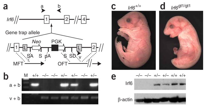
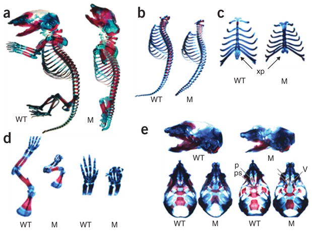
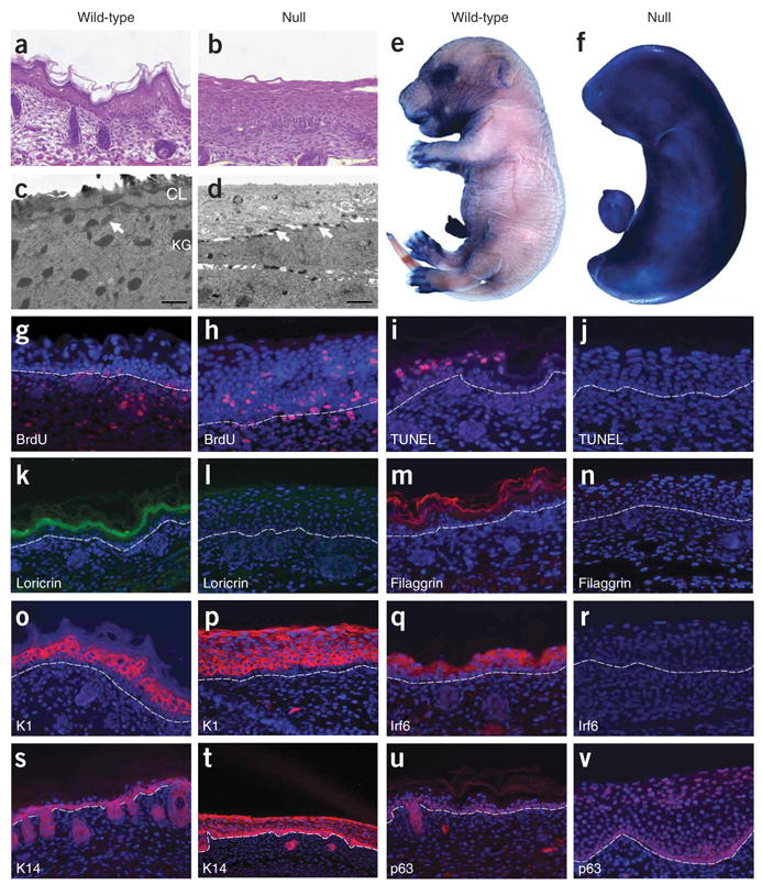
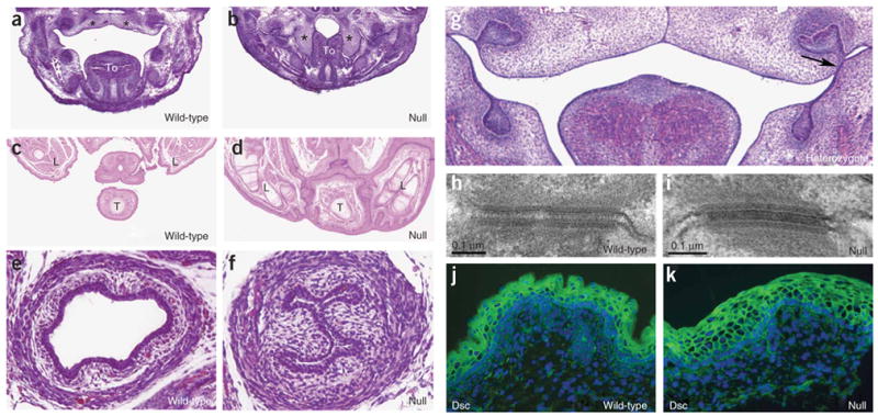
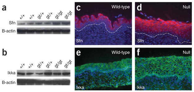
Transcription factor paralogs may share a common role in staged or overlapping expression in specific tissues, as in the Hox family. In other cases, family members have distinct roles in a range of embryologic, differentiation or response pathways (as in the Tbx and Pax families). For the interferon regulatory factor (IRF) family of transcription factors, mice deficient in Irf1, Irf2, Irf3, Irf4, Irf5, Irf7, Irf8 or Irf9 have defects in the immune response but show no embryologic abnormalities. Mice deficient for Irf6 have not been reported, but in humans, mutations in IRF6 cause two mendelian orofacial clefting syndromes, and genetic variation in IRF6 confers risk for isolated cleft lip and palate. Here we report that mice deficient for Irf6 have abnormal skin, limb and craniofacial development. Histological and gene expression analyses indicate that the primary defect is in keratinocyte differentiation and proliferation. This study describes a new role for an IRF family member in epidermal development.


Figures

Figure 1
Figure 1
Genotypic and phenotypic analysis of mice deficient for Irf6. (a) Irf6 gene trap allele (Irf6gt1). The gene trap vector (VICTR48) inserted 36 bp from the splice donor site of intron 1 and contains flanking long terminal repeats (L), a splice acceptor (SA) and donor (SD) sites, the ORF for neomycin (Neo), stop codons (S), polyadenylation site (pA), the phosphoglycerate kinase (PGK) promoter, marker fusion transcript (MFT) and OmniBank sequence tag (OST) fusion transcript (OFT). (b) Genotypic analysis by PCR of genomic DNA derived from E17.5 embryos using primers a and b to detect wild-type (+) and using primers v and b to detect mutant (−) alleles. (c) Gross appearance of wild-type E17.5 embryos. (d) E17.5 embryo that is homozygous for gene trap allele (Irf6gt1/gt1). (e) Protein blot analysis of protein extracts obtained from E17.5 skin from mice with the indicated genotype. Blots were probed with antibodies directed against the indicated protein.
Figure 2
Figure 2
Skeletal defects in mice deficient for Irf6. (a) Skeletal structures of E17.5 wild-type (WT) and newborn mutant (M) embryos were stained for bone (alizarin red S) and cartilage (alcian blue). (b) The spine showed equal numbers of vertebrae in wild-type and null embryos. However, vertebrae in the mutant appeared smaller and showed delayed ossification. The mutant tail was substantially shorter. (c) There were equal numbers of ribs in the wild-type and null embryos, but the sternum appeared shorter with delayed ossification. The xiphoid process (xp) was bifid in the null embryo, demonstrating a failure of complete fusion of the thoracic cage. (d) The long bones were slightly shorter in null versus wild-type littermates, but both null and wild-type embryos showed normal surrounding muscle when skin was dissected. Proximal bones of the upper and lower limb were present in the null embryos, but distal structures, notably the digits, were severely abnormal. Forepaws were magnified to display synostosis of digits and absence of distal phalanges. (e) Skulls from wild-type and null embryos in lateral (top), superior (bottom left) and inferior (bottom right) views. Mandibles were removed for inferior views. The mandible in the null embryo was smaller with a narrower angle than in the wild-type, and the snout was also shorter in the null embryo. The palate shelves (p) in the wild-type embryo fuse along the midline, but the null embryo has a cleft palate. The palate shelves of null embryo are posteriorly and laterally displaced, allowing for direct viewing of nasal structures, including the vomer (v). The presphenoid (ps) is absent in the mutant. Similar results were obtained from three skeletal preparations.
Figure 3
Figure 3
Histologic and molecular analyses of Irf6-null E17.5 embryos. (ad) Hematoxylin and eosin–stained sections (a,b) and electron micrographs (c,d) of back skin. Note the presence of keratohyalin granules (KG) in the granular layer and a cornified layer (CL) of wild-type epidermis (c), which are absent in null epidermis (d). Arrows indicate desmosomes. Dye exclusion assays of whole embryos show a functional barrier in wild-type (e) but not in null (f) embryos. Cellular proliferation and programmed cell death were evaluated by BrdU (g,h) and TUNEL (i,j) staining, respectively. Immunofluorescent staining (red, except loricrin is green) for cornified and granular layers (km) and for spinous (or) and basal (sv) layers. Proteins against which antibodies were directed are indicated. Nuclei were counterstained with DAPI (blue), and the basement membrane is indicated by the dashed line.
Figure 4
Figure 4
Epidermal adhesions and desmosomal structure in Irf6-null embryos. (af) Hematoxylin and eosin staining for sections of E14.5 frontal head (a,b), E17.5 rostral trunk (c,d) and E17.5 caudal trunk (e,f). Palate shelves elevate and begin to fuse in wild-type mice (a) but not in null embryos (b), where adhesions contribute to crowding of the oral cavity and prevent the palate shelves from elevating. Hindlimbs and tail are joined by epithelial adhesions (compare c with d). Esophagus is closed in null embryo (compare e with f). *: palate shelves; To: tongue; L: hindlimb; T: tail. Adhesion in the heterozygote was observed superficial to the first molar on the left side only (g, arrow). (h,i) Electron micrographs of desmosomes. Immunofluorescent staining (green) for desmocollin (Dsc) (j,k). We did not observe any difference in desmosomal structure and protein between wild-type and null embryos. Nuclei were counterstained with DAPI (blue).
Figure 5
Figure 5
Expression of stratifin and Ikka in skin from Irf6-null E17.5 embryos. (a,b) Protein blot analyses show that stratifin expression (a) is greater in null embryos, whereas Ikka expression (b) does not seem to change. The respective blots were stripped and reprobed with β-actin as a control for loading. (c) Immunostaining shows that stratifin is expressed in the cytoplasm of spinous cells in skin from wild-type E17.5 embryos (c); it remains cytoplasmic but increases along with the expanded spinous layer in the skin from null embryos (d). (e,f) Immunostaining shows that Ikka is expressed predominantly in the basal layer in skin from both the wild-type (e) and Irf6-null (f) embryos. Ikka is localized to the cytoplasm and in the nucleus of some cells. This distribution does not appear to differ in skin from wild-type and Irf6-null embryos.

References

    1. Matsuyama T, et al. Targeted disruption of IRF-1 or IRF-2 results in abnormal type I IFN gene induction and aberrant lymphocyte development. Cell. 1993;75:83–97. - PubMed
    1. Sato M, et al. Distinct and essential roles of transcription factors IRF-3 and IRF-7 in response to viruses for IFN-alpha/beta gene induction. Immunity. 2000;13:539–548. - PubMed
    1. Mittrucker HW, et al. Requirement for the transcription factor LSIRF/IRF4 for mature B and T lymphocyte function. Science. 1997;275:540–543. - PubMed
    1. Takaoka A, et al. Integral role of IRF-5 in the gene induction programme activated by Toll-like receptors. Nature. 2005;434:243–249. - PubMed
    1. Honda K, et al. IRF-7 is the master regulator of type-I interferon-dependent immune responses. Nature. 2005;434:772–777. - PubMed

Publication types

MeSH terms

Substances

Associated data