Case Reports
. 2006 Nov;27(11):1157.
doi: 10.1002/humu.9461.

Recessive arrhythmogenic right ventricular dysplasia due to novel cryptic splice mutation in PKP2

Affiliations
Case Reports

Recessive arrhythmogenic right ventricular dysplasia due to novel cryptic splice mutation in PKP2

Mark M Awad et al. Hum Mutat. 2006 Nov.

Abstract

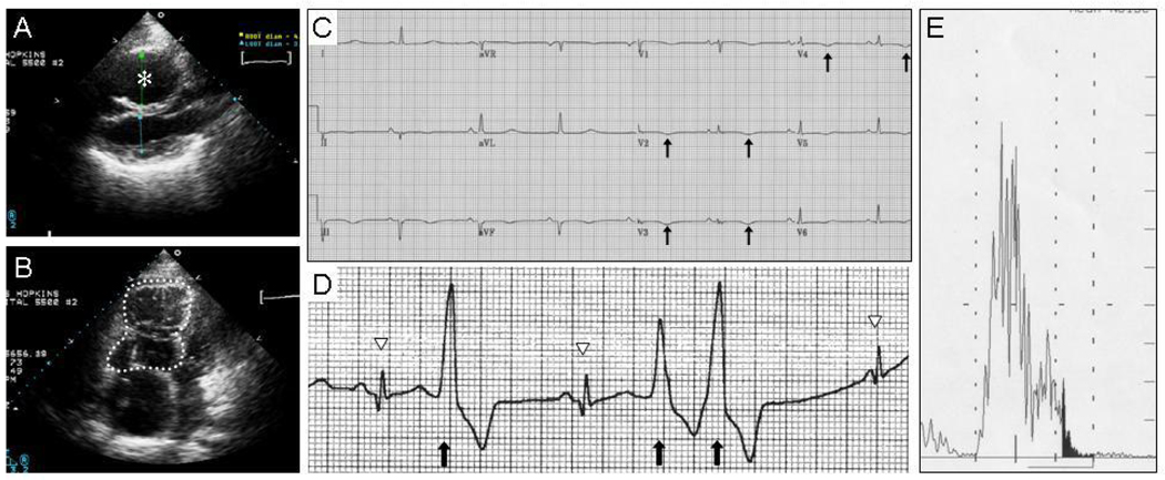
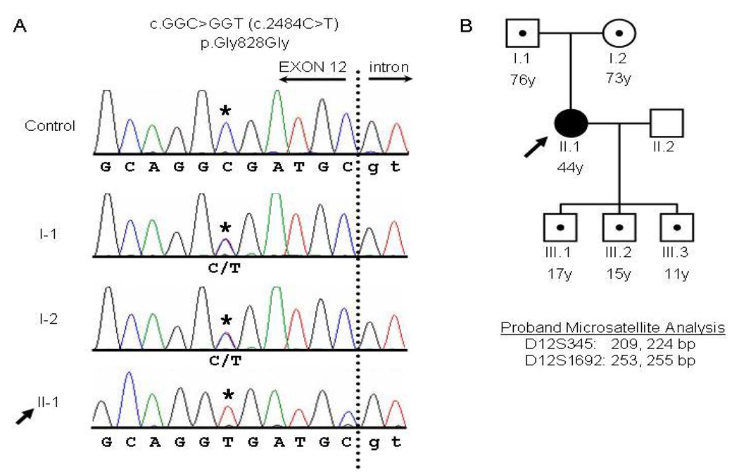
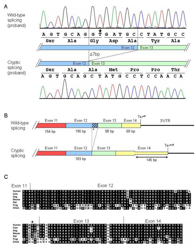
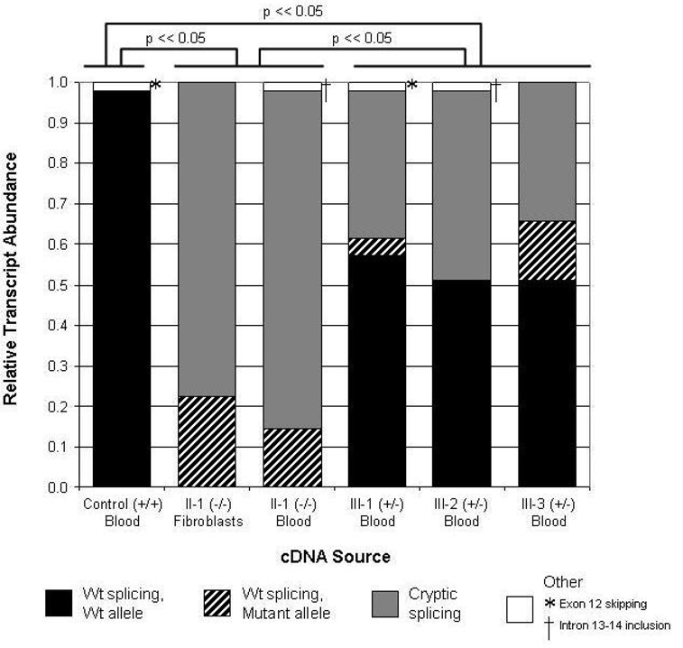
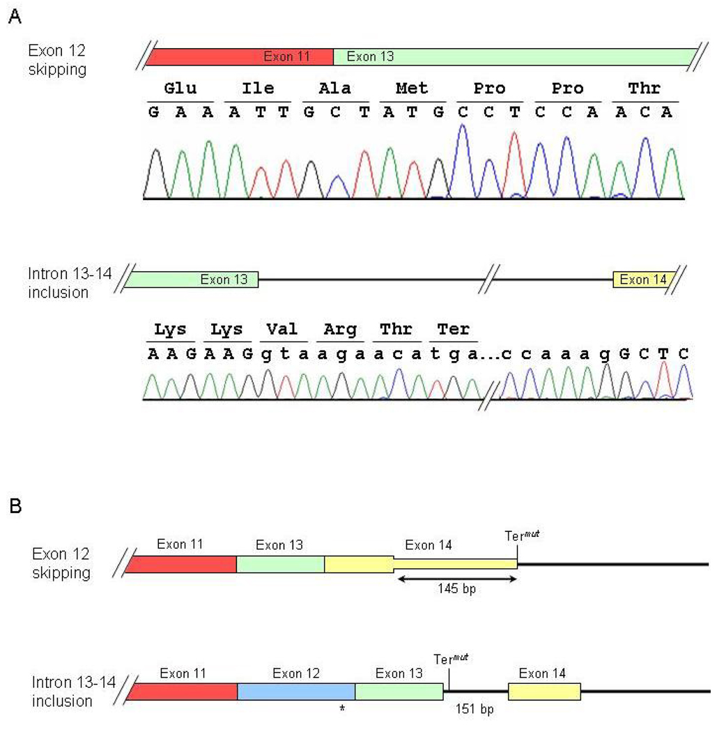
Arrhythmogenic right ventricular dysplasia (ARVD) is a genetic disorder resulting in fibro-fatty replacement of right ventricular myocytes and consequent ventricular arrhythmias. Heterozygous mutations in PKP2 encoding plakophilin-2 have previously been reported to cause dominant ARVD with reduced penetrance. We report the first case of recessive ARVD caused by mutations in PKP2. Candidate gene analysis in a typical proband with this disorder identified a novel homozygous mutation in PKP2 (c.[2484C>T]+[2484C>T]), which is predicted to be translationally silent (p.Gly828). Analysis of the proband's mRNA, however, shows that this mutation causes predominantly cryptic splicing, with a 7-nucleotide deletion in exon 12. The ensuing frame shift disrupts the last 54 amino acids of plakophilin-2 and extends the open reading frame by 145 nucleotides (48 amino acids) into the 3' untranslated region. Haplotype analysis demonstrates the absence of remote consanguinity. Heterozygous family members produce approximately 60% of properly spliced PKP2 and do not have manifestations of ARVD. Further analysis of PKP2 mRNA sequence revealed two additional alternatively spliced transcripts. The possibility of cryptic or alternative splicing should be considered with identification of apparently synonymous nucleotide substitutions in this gene.


Figures

Figure 1
Figure 1
Proband meets diagnostic criteria for ARVD. A, B: Right ventricular dilatation on echocardiogram, shown from a parasternal long axis view (A), and from an apical four-chamber view (B). The right ventricle is marked with an asterisk in (A) and is traced with a dotted line in (B). C: T-wave inversions (arrows) in precordial leads V2 to V4) on 12-lead electrocardiogram (ECG). D: Premature ventricular contractions (PVCs, arrows) on Holter monitoring. Normal sinus beats are denoted with arrowheads. E: Late potentials (shaded region) on signal averaged ECG.
Figure 2
Figure 2
PKP2 genomic sequencing and pedigree analysis. A: PKP2 genomic sequence analysis of the end of exon 12 and the downstream 5’ intronic splice site for a control individual, the proband (II-1), and the proband’s parents (I-1 and I-2). The dotted line indicates the boundary between the exon (capital letters) and intron (lowercase). The c.2484C>T mutation (NM_004572.2) is indicated with an asterisk. The proband’s parents are each heterozygous (C/T) at position c.2484, and the proband (arrow) is homozygous (T/T) at this position. B: Pedigree of the proband’s family. Squares, male; circles, female; black shading, affected; white shading, unaffected; dots indicate heterozygous individuals as described in (A). For the proband, the lengths of the microsatellite markers D12S345 and D12S1692 are indicated below the pedigree.
Figure 3
Figure 3
Cryptic splicing of the PKP2 transcript caused by the 2484C→T mutation. A: Chromatograms of cloned RT-PCR products. Top panel: wild-type splicing in the proband. Bottom panel: mutant splicing results in deletion of the last seven nucleotides in exon 12 and causes a subsequent frame shift. B: Diagram illustrating consequences of cryptic splicing on translation. Exons are indicated by boxes, and the 3’UTR is drawn as a line. Terwt and Termut indicate the wild-type and mutant termination codons, respectively. C: ClustalW sequence alignment of plakophilin-2 orthologs demonstrating conservation of the C-terminal residues that are disrupted in the mutant splice forms. The G828G mutation is indicated with an asterisk (*). Black shading, residue identity; gray shading, conservation of residue character.
Figure 4
Figure 4
Quantification of wild-type and mutant cryptic splicing of the PKP2 transcript in six clinical samples. Relative transcript abundance in two sources of RNA from the proband is compared to transcript abundance in RNA derived from an unaffected control and from the proband’s three heterozygous offspring. Proportion of wild-type splicing derived from the wild-type allele (c.2484C) is shaded in black. Proportion of wild-type splicing derived from the mutant allele (c.2484T) is shaded with black diagonal bars. Proportion of cryptic splicing with the 7-bp deletion at the end of exon 12 is shaded in gray. Minor splice forms (see Fig. 5), shaded in white, include exon 12 skipping (*) and intron 13–14 retention (†). Statistical differences in the relative abundance of wild-type and mutant splice forms are indicated above the graph, comparing control, combined proband samples, and combined heterozygous samples.
Figure 5
Figure 5
Two novel splice forms of PKP2. A: Chromatograms of RT-PCR products demonstrating skipping of exon 12 (top panel) and retention of intron 13–14 (bottom panel). B: Diagram illustrating consequences of alternative splice forms on translation. Exons are indicated by boxes; the intron and 3’UTR is drawn as lines. Termut indicates the mutant termination codon.

References

    1. Alcalai R, Metzger S, Rosenheck S, Meiner V, Chajek-Shaul T. A recessive mutation in desmoplakin causes arrhythmogenic right ventricular dysplasia, skin disorder, and woolly hair. J Am Coll Cardiol. 2003;42(2):319–327. - PubMed
    1. Awad MM, Dalal D, Cho E, Amat-Alarcon N, James C, Tichnell C, Tucker A, Russell SD, Bluemke DA, Dietz HC, et al. DSG2 Mutations Contribute to Arrhythmogenic Right Ventricular Dysplasia/Cardiomyopathy. Am J Hum Genet. 2006;79:136–142. - PMC - PubMed
    1. Bauce B, Basso C, Rampazzo A, Beffagna G, Daliento L, Frigo G, Malacrida S, Settimo L, Danieli G, Thiene G, et al. Clinical profile of four families with arrhythmogenic right ventricular cardiomyopathy caused by dominant desmoplakin mutations. Eur Heart J. 2005;26(16):1666–1675. - PubMed
    1. Beffagna G, Occhi G, Nava A, Vitiello L, Ditadi A, Basso C, Bauce B, Carraro G, Thiene G, Towbin JA, et al. Regulatory mutations in transforming growth factor-beta3 gene cause arrhythmogenic right ventricular cardiomyopathy type 1. Cardiovasc Res. 2005;65(2):366–373. - PubMed
    1. Chen X, Bonne S, Hatzfeld M, van Roy F, Green KJ. Protein binding and functional characterization of plakophilin 2. Evidence for its diverse roles in desmosomes and beta -catenin signaling. J Biol Chem. 2002;277(12):10512–10522. - PubMed

Publication types

LinkOut - more resources