. 2006 Oct 16:7:36.
doi: 10.1186/1471-2199-7-36.

Expression and loss of alleles in cultured mouse embryonic fibroblasts and stem cells carrying allelic fluorescent protein genes

Affiliations

Expression and loss of alleles in cultured mouse embryonic fibroblasts and stem cells carrying allelic fluorescent protein genes

Jon S Larson et al. BMC Mol Biol. .

Abstract

Background: Loss of heterozygosity (LOH) contributes to many cancers, but the rate at which these events occur in normal cells of the body is not clear. LOH would be detectable in diverse cell types in the body if this event were to confer an obvious cellular phenotype. Mice that carry two different fluorescent protein genes as alleles of a locus would seem to be a useful tool for addressing this issue because LOH would change a cell's phenotype from dichromatic to monochromatic. In addition, LOH caused by mitotic crossing over might be discernable in tissues because this event produces a pair of neighboring monochromatic cells that are different colors.

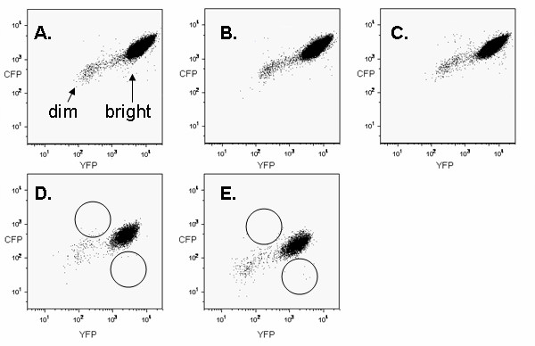
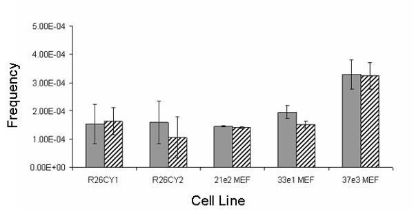
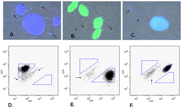
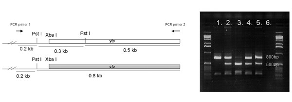
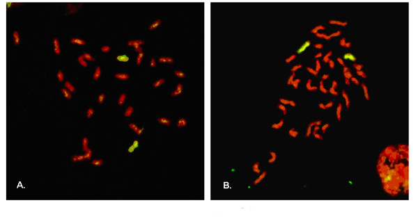
Results: As a step in assessing the utility of this approach, we derived primary embryonic fibroblast populations and embryonic stem cell lines from mice that carried two different fluorescent protein genes as alleles at the chromosome 6 locus, ROSA26. Fluorescence activated cell sorting (FACS) showed that the vast majority of cells in each line expressed the two marker proteins at similar levels, and that populations exhibited expression noise similar to that seen in bacteria and yeast. Cells with a monochromatic phenotype were present at frequencies on the order of 10(-4) and appeared to be produced at a rate of approximately 10(-5) variant cells per mitosis. 45 of 45 stably monochromatic ES cell clones exhibited loss of the expected allele at the ROSA26 locus. More than half of these clones retained heterozygosity at a locus between ROSA26 and the centromere. Other clones exhibited LOH near the centromere, but were disomic for chromosome 6.

Conclusion: Allelic fluorescent markers allowed LOH at the ROSA26 locus to be detected by FACS. LOH at this locus was usually not accompanied by LOH near the centromere, suggesting that mitotic recombination was the major cause of ROSA26 LOH. Dichromatic mouse embryonic cells provide a novel system for studying genetic/karyotypic stability and factors influencing expression from allelic genes. Similar approaches will allow these phenomena to be studied in tissues.


Figures

Figure 1
Figure 1
FACS dot plots (106 events) showing fluorescent profiles of three R26CY MEF cell lines (A-C) and two ES cell lines (D, E). Arrows in panel A indicate "dim" and "bright" events in MEF plots. Circles in panels D and E indicate areas where monochromatic variants would be expected to be located.
Figure 2
Figure 2
Expression of CFP (A) and YFP (B) in a mouse embryo (14.5 dpc), heterozygous at the ROSA26 locus. Panel C shows an image produced by overlaying the CFP and YFP signals.
Figure 3
Figure 3
Variation in fluorescent signals (noise) from 1000 randomly selected cells. (A) MEF cell line 33e1 (B) ES cell line R26CY1. The fluorescent units are relative values assigned by the sorter. Intensities for CFP have been normalized to correct for its relative faintness when compared to YFP intensity. Arrows at right angles indicate noise axes, extrinsic (E) and intrinsic (I).
Figure 4
Figure 4
Abundance of spontaneously occurring apparent monochromatic variants. Solid and hatched bars indicate frequencies of cells exhibiting only CFP or only YFP, respectively. R26CY1 and R26CY2 are data from the two ES cells lines. 21e2MEF, 33e1MEF and 37e3MEF are data from the three MEF lines. Error bars indicate the standard errors of the means.
Figure 5
Figure 5
Phenotypic analysis of FACS-isolated ES cell clones. Panels A-C show images of three clones with different phenotypes. (A) colony of cells expressing CFP only, (B) colony of cells expressing YFP only, (C) colony of cells expressing both CFP and YFP. Images shown were are produced by merging three images, CFP epifluorescence, YFP epifluorescence, and phase contrast. Gray cells under the ES clones are a monolayer of non-fluorescent wild type MEF feeder cells. Arrows indicate some of the cellular debris and dead cells that exhibited autofluorescence. Panels D, E and F correlate with panels A, B and C, respectively, and show FACS analysis of each subclone. Polygons indicate gates used to collect putative monochromatic cells. Arrows indicate residual MEF feeder cells present in ES cell populations subjected to FACS.
Figure 6
Figure 6
Genotyping the ROSA26 locus in DNA isolated from monochromatic clones. At left are shown maps of the two alleles in parental cells. The YFP gene has an additional cleavage site for Pst1 restriction endonuclease. Lane 1, a clone lacking the YFP gene. Lane 2, a dichromatic clone, which retained both the genes, as expected. Lane 3, a clone lacking the CFP gene. Lanes 4 & 5, data from DNA of unsorted ES cell lines. Lane 6, PCR control containing no DNA in the reaction mix. Markers are 1 Kb DNA ladder (Invitrogen).
Figure 7
Figure 7
Whole chromosome-6-paint FISH analysis of monochromatic ES cell clones that had lost heterozygosity at both the ROSA26 locus and D6Mit159. Metaphase chromosomes were hybridized to a chromosome 6 probe labeled with FITC. Chromosomes were counter stained red with propidium iodide (PI). Images shown were produced by merging FITC and PI epifluorescence images. Copies of chromosome 6 are yellow. Other chromosomes are red. Panels A and B show metaphase chromosomes from cells that expressed CFP only or YFP only, respectively. 400× magnification.

Similar articles

Cited by

References

    1. Scrable HJ, Witte DP, Lampkin BC, Cavenee WK. Chromosomal localization of the human rhabdomyosarcoma locus by mitotic recombination mapping. Nature. 1987;329:645–647. doi: 10.1038/329645a0. - DOI - PubMed
    1. Cavenee WK, Dryja TP, Phillips RA, Benedict WF, Godbout R, Gallie BL, Murphree AL, Strong LC, White RL. Expression of recessive alleles by chromosomal mechanisms in retinoblastoma. Nature. 1983;305:779–784. doi: 10.1038/305779a0. - DOI - PubMed
    1. Cavenee WK. The recessive nature of dominance. Genes Chromosomes Cancer. 2003;38:322–325. doi: 10.1002/gcc.10253. - DOI - PubMed
    1. Knudson AG. Cancer genetics. Am J Med Genet. 2002;111:96–102. doi: 10.1002/ajmg.10320. - DOI - PubMed
    1. Lefebvre L, Dionne N, Karaskova J, Squire JA, Nagy A. Selection for transgene homozygosity in embryonic stem cells results in extensive loss of heterozygosity. Nat Genet. 2001;27:257–258. doi: 10.1038/85808. - DOI - PubMed

Publication types

MeSH terms