

Atypically diffuse functional connectivity between caudate nuclei and cerebral cortex in autism
- PMID: 17042953
- PMCID: PMC1635430
- DOI: 10.1186/1744-9081-2-34
Atypically diffuse functional connectivity between caudate nuclei and cerebral cortex in autism
Abstract
Background: Autism is a neurodevelopmental disorder affecting sociocommunicative behavior, but also sensorimotor skill learning, oculomotor control, and executive functioning. Some of these impairments may be related to abnormalities of the caudate nuclei, which have been reported for autism.
Methods: Our sample was comprised of 8 high-functioning males with autism and 8 handedness, sex, and age-matched controls. Subjects underwent functional MRI scanning during performance on simple visuomotor coordination tasks. Functional connectivity MRI (fcMRI) effects were identified as interregional blood oxygenation level dependent (BOLD) signal cross-correlation, using the caudate nuclei as seed volumes.
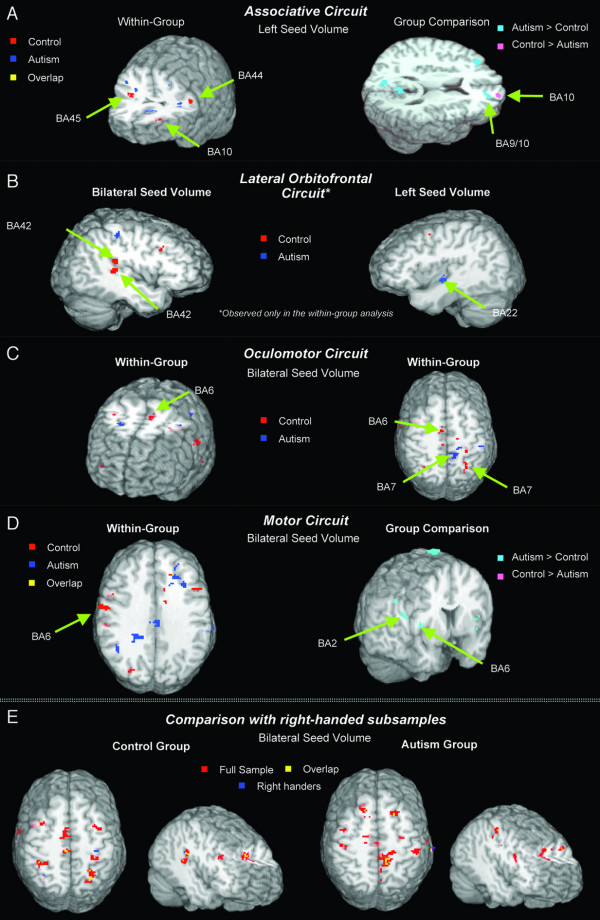
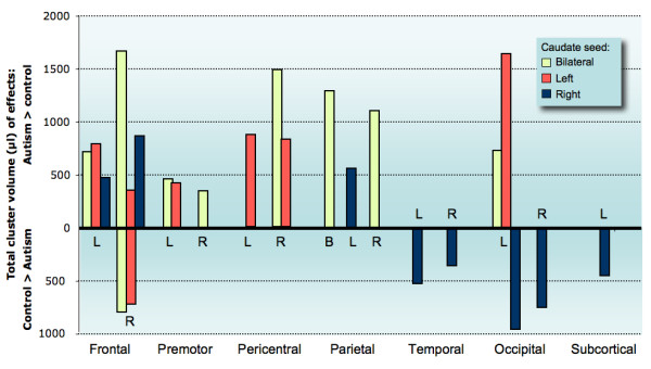
Results: In the control group, fcMRI effects were found in circuits with known participation of the caudate nuclei (associative, orbitofrontal, oculomotor, motor circuits). Although in the autism group fcMRI effects within these circuits were less pronounced or absent, autistic subjects showed diffusely increased connectivity mostly in pericentral regions, but also in brain areas outside expected anatomical circuits (such as visual cortex).
Conclusion: These atypical connectivity patterns may be linked to developmental brain growth disturbances recently reported in autism and suggest inefficiently organized functional connectivity between caudate nuclei and cerebral cortex, potentially accounting for stereotypic behaviors and executive impairments.
Figures
Similar articles
-
Partially enhanced thalamocortical functional connectivity in autism.Brain Res. 2006 Aug 9;1104(1):160-74. doi: 10.1016/j.brainres.2006.05.064. Epub 2006 Jul 7. Brain Res. 2006. PMID: 16828063
-
Altered corticostriatal functional connectivity in obsessive-compulsive disorder.Arch Gen Psychiatry. 2009 Nov;66(11):1189-200. doi: 10.1001/archgenpsychiatry.2009.152. Arch Gen Psychiatry. 2009. PMID: 19884607
-
Abnormal activity patterns in premotor cortex during sequence learning in autistic patients.Biol Psychiatry. 2004 Sep 1;56(5):323-32. doi: 10.1016/j.biopsych.2004.06.007. Biol Psychiatry. 2004. PMID: 15336514
-
Brain asymmetries in autism and developmental language disorder: a nested whole-brain analysis.Brain. 2005 Jan;128(Pt 1):213-26. doi: 10.1093/brain/awh330. Epub 2004 Nov 24. Brain. 2005. PMID: 15563515
-
Regional gray matter volumetric changes in autism associated with social and repetitive behavior symptoms.BMC Psychiatry. 2006 Dec 13;6:56. doi: 10.1186/1471-244X-6-56. BMC Psychiatry. 2006. PMID: 17166273 Free PMC article.
Cited by
-
Functional connectivity-based subtypes of individuals with and without autism spectrum disorder.Netw Neurosci. 2019 Feb 1;3(2):344-362. doi: 10.1162/netn_a_00067. eCollection 2019. Netw Neurosci. 2019. PMID: 30793086 Free PMC article.
-
Abnormalities in fronto-striatal connectivity within language networks relate to differences in grey-matter heterogeneity in Asperger syndrome.Neuroimage Clin. 2013 May 27;2:716-26. doi: 10.1016/j.nicl.2013.05.010. eCollection 2013. Neuroimage Clin. 2013. PMID: 24179823 Free PMC article.
-
Autism Spectrum Disorders and Drug Addiction: Common Pathways, Common Molecules, Distinct Disorders?Front Neurosci. 2016 Feb 5;10:20. doi: 10.3389/fnins.2016.00020. eCollection 2016. Front Neurosci. 2016. PMID: 26903789 Free PMC article. Review.
-
Repetitive behaviors in autism are linked to imbalance of corticostriatal connectivity: a functional connectivity MRI study.Soc Cogn Affect Neurosci. 2018 Jan 1;13(1):32-42. doi: 10.1093/scan/nsx129. Soc Cogn Affect Neurosci. 2018. PMID: 29177509 Free PMC article.
-
Temporal fractal analysis of the rs-BOLD signal identifies brain abnormalities in autism spectrum disorder.PLoS One. 2017 Dec 22;12(12):e0190081. doi: 10.1371/journal.pone.0190081. eCollection 2017. PLoS One. 2017. PMID: 29272297 Free PMC article.
References
-
- Minshew NJ, Goldstein G, Siegel DJ. Neuropsychologic functioning in autism: Profile of a complex information processing disorder. J Int Neuropsychol Soc. 1997;3:303–316. - PubMed
Grants and funding
LinkOut - more resources
Full Text Sources
Other Literature Sources
