. 2006;8(6):R159.
doi: 10.1186/ar2066.

Alterations of metabolic activity in human osteoarthritic osteoblasts by lipid peroxidation end product 4-hydroxynonenal

Affiliations

Alterations of metabolic activity in human osteoarthritic osteoblasts by lipid peroxidation end product 4-hydroxynonenal

Qin Shi et al. Arthritis Res Ther. 2006.

Abstract

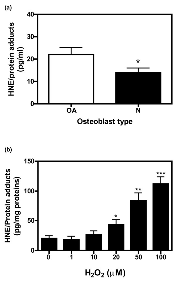
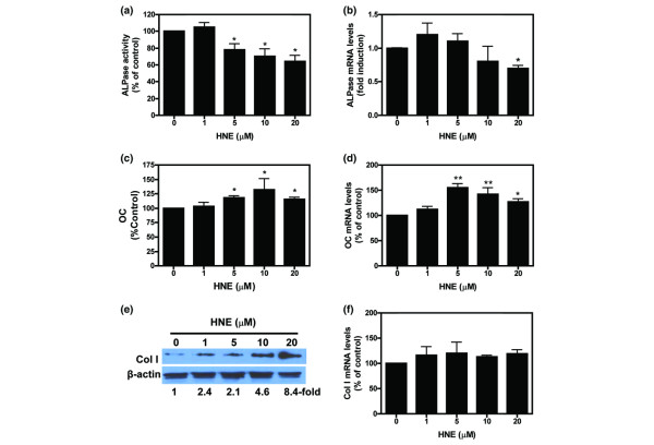
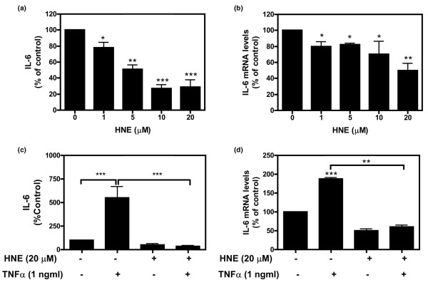
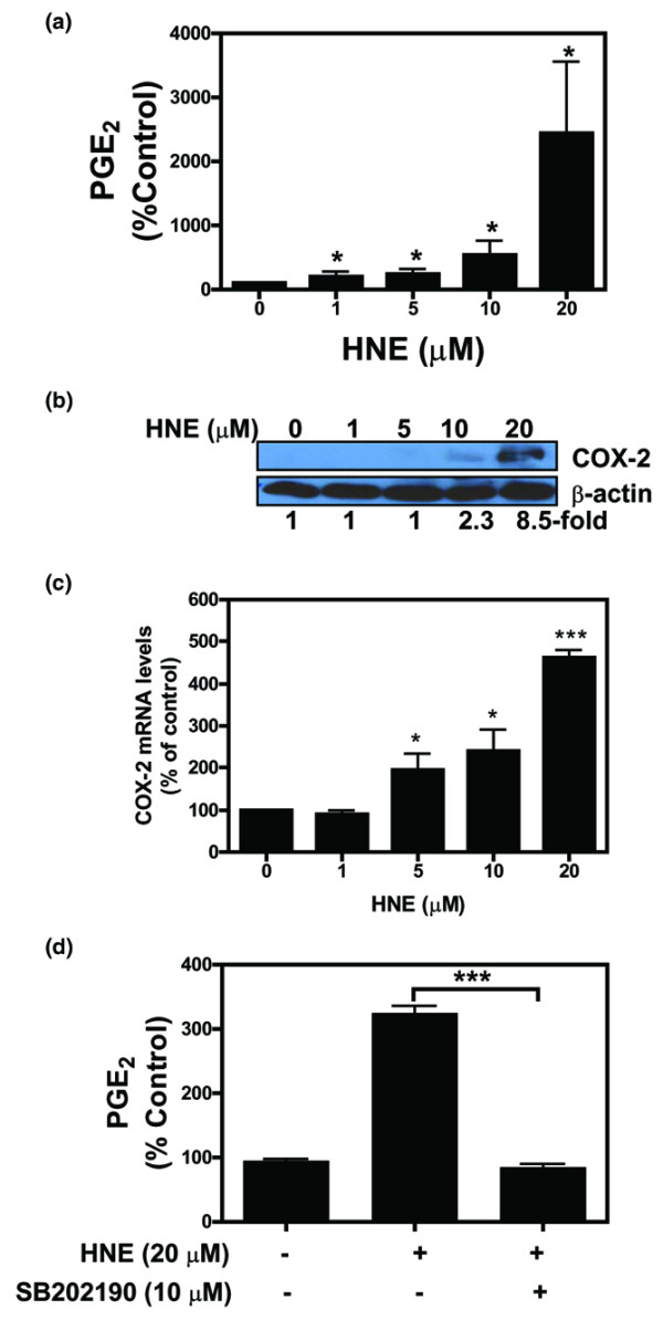
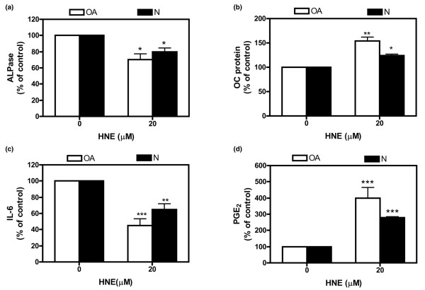
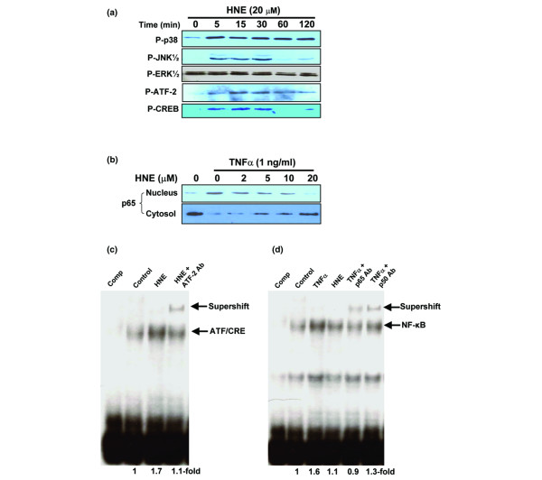
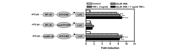
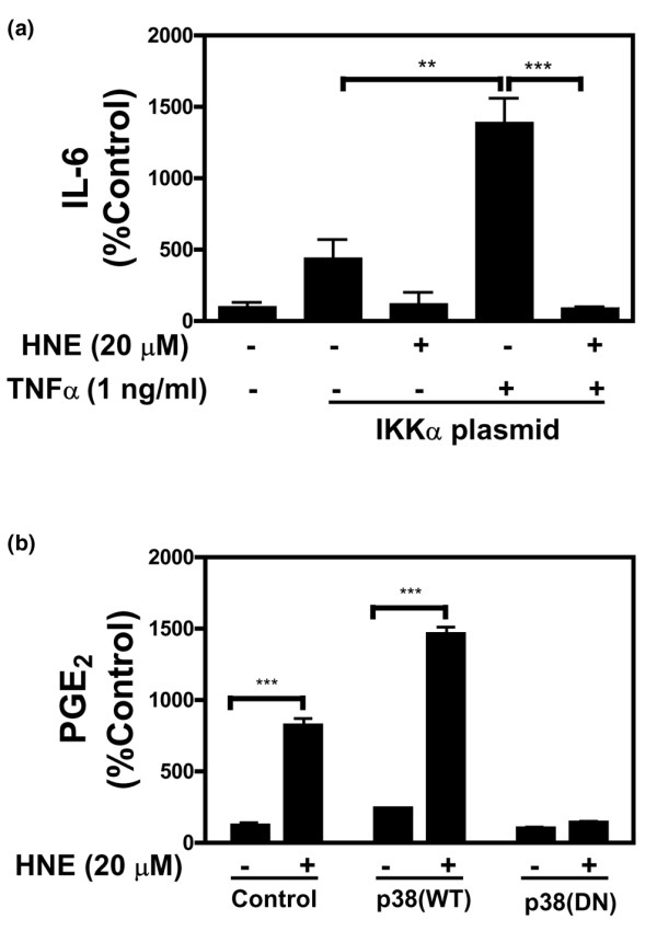
4-Hydroxynonenal (HNE), a lipid peroxidation end product, is produced abundantly in osteoarthritic (OA) articular tissues, but its role in bone metabolism is ill-defined. In this study, we tested the hypothesis that alterations in OA osteoblast metabolism are attributed, in part, to increased levels of HNE. Our data showed that HNE/protein adduct levels were higher in OA osteoblasts compared to normal and when OA osteoblasts were treated with H2O2. Investigating osteoblast markers, we found that HNE increased osteocalcin and type I collagen synthesis but inhibited alkaline phosphatase activity. We next examined the effects of HNE on the signaling pathways controlling cyclooxygenase-2 (COX-2) and interleukin-6 (IL-6) expression in view of their putative role in OA pathophysiology. HNE dose-dependently decreased basal and tumour necrosis factor-alpha (TNF-alpha)-induced IL-6 expression while inducing COX-2 expression and prostaglandin E2 (PGE2) release. In a similar pattern, HNE induces changes in osteoblast markers as well as PGE2 and IL-6 release in normal osteoblasts. Upon examination of signaling pathways involved in PGE2 and IL-6 production, we found that HNE-induced PGE2 release was abrogated by SB202190, a p38 mitogen-activated protein kinase (MAPK) inhibitor. Overexpression of p38 MAPK enhanced HNE-induced PGE2 release. In this connection, HNE markedly increased the phosphorylation of p38 MAPK, JNK2, and transcription factors (CREB-1, ATF-2) with a concomitant increase in the DNA-binding activity of CRE/ATF. Transfection experiments with a human COX-2 promoter construct revealed that the CRE element (-58/-53 bp) was essential for HNE-induced COX-2 promoter activity. However, HNE inhibited the phosphorylation of IkappaBalpha and subsequently the DNA-binding activity of nuclear factor-kappaB. Overexpression of IKKalpha increased TNF-alpha-induced IL-6 production. This induction was inhibited when TNF-alpha was combined with HNE. These findings suggest that HNE may exert multiple effects on human OA osteoblasts by selective activation of signal transduction pathways and alteration of osteoblastic phenotype expression and pro-inflammatory mediator production.


Figures

Figure 1
Figure 1
Determination of HNE/protein adduct concentrations in normal (N) and osteoarthritic (OA) osteoblast. HNE/protein adduct levels were measured by enzyme-linked immunosorbent assay in cellular extracts from untreated (a) or treated (b) osteoblasts with increasing concentrations of H2O2 for 24 hours at the indicated concentrations. HNE/protein adduct levels were expressed in picograms of HNE/protein adducts per milligrams of total proteins. Data are mean ± standard error of the mean (n = 3). Statistics: Student unpaired t test; *p < 0.05, **P < 0.01, ***P < 0.001. HNE, 4-hydroxynonenal.
Figure 2
Figure 2
Effect of HNE on osteoblast markers ALPase, OC, and Col I. Human osteoarthritic osteoblasts were incubated with increasing concentrations of HNE for 48 hours and then ALPase activity (a) and Col I protein level (e) were determined in cellular extract as described in Materials and methods. The OC release (c) was determined in culture medium by enzyme-linked immunosorbent assay. For mRNA level, cells were incubated for 4 hours in the absence or presence of indicated concentrations of HNE, total RNA was isolated and reverse-transcribed into cDNA, and ALPase (b), OC (d), and Col I (f) were quantified using real-time polymerase chain reaction. All experiments were performed in triplicate, and negative controls without template RNA were included in each experiment as indicated in Materials and methods. mRNA levels were normalised to those of GAPDH (glyceraldehyde-3-phosphate dehydrogenase) mRNA. Data are means ± standard error of the mean of n = 3 and expressed as a percentage of untreated cells. Statistics: Student unpaired t test; *p < 0.05, **p < 0.01. ALPase, alkaline phosphatase; Col I, type I collagen; HNE, 4-hydroxynonenal; OC, osteocalcin.
Figure 3
Figure 3
Effect of HNE on IL-6 protein production in osteoblasts. Osteoblasts were treated with HNE (0 to 20 μM) for 48 or 4 hours for IL-6 protein (a) (n = 7) and mRNA (b) (n = 3) determination, respectively. The effect of HNE combined with TNF-α was evaluated by incubating osteoblasts with HNE (20 μM) for 30 minutes and subsequently stimulating them with TNF-α (1 ng/ml) for 48 or 4 hours for IL-6 protein (c) (n = 7) and mRNA (d) (n = 3) determination, respectively. mRNA levels of each gene were quantified by real-time polymerase chain reaction as described in Materials and methods and normalised to those of GAPDH (glyceraldehyde-3-phosphate dehydrogenase) mRNA. Data are means ± standard error of the mean and expressed as a percentage of untreated cells. Statistics: Student unpaired t test; *p < 0.05, **p < 0.01, ***p < 0.001. HNE, 4-hydroxynonenal; IL-6, interleukin-6; TNF-α, tumour necrosis factor-α.
Figure 4
Figure 4
Effect of HNE on PGE2 release and COX-2 expression.(a) Osteoblasts were treated with HNE (0 to 20 μM) for 48 hours, and PGE2 release was evaluated in culture medium by PGE2 enzyme immunoassay kit. (b, c) Osteoblasts were treated with HNE (0 to 20 μM) for 48 or 4 hours for protein and mRNA determination, respectively. COX-2 protein expression (b) and mRNA expression (c) were evaluated by Western blot and real-time reverse transcriptase-polymerase chain reaction, respectively. Quantifications of COX-2 protein and mRNA levels were normalised, respectively, to those of β-actin protein and GAPDH (glyceraldehyde-3-phosphate dehydrogenase) mRNA. (d) Cells were preincubated in the absence or presence of p38 MAPK inhibitor SB202190 (10 μM) for 30 minutes, followed by incubation by HNE (20 μM) for 48 hours. PGE2 secretion was evaluated as described above. Data are means ± standard error of the mean of n = 3 and expressed as a percentage of untreated cells. Statistics: Student unpaired t test; *p < 0.05, ***p < 0.001. COX-2, cyclooxygenase-2; HNE, 4-hydroxynonenal; MAPK, mitogen-activated protein kinase; PGE2, prostaglandin E2.
Figure 5
Figure 5
Comparison of the effect of HNE on normal (N) and osteoarthtitic (OA) osteoblast metabolism. Cells were incubated in the absence or presence of 20 μM HNE for 48 hours. ALPase activity (a) was determined in cellular extract as described in Materials and methods. OC (b), IL-6 (c), and PGE2 (d) levels were measured in culture media using specific kits. Data are means ± standard error of the mean of n = 3 and expressed as a percentage of untreated cells. Statistics: Student unpaired t test; *p < 0.05, **p < 0.01, ***p < 0.001. ALPase, alkaline phosphatase; HNE, 4-hydroxynonenal; IL-6, interleukin-6; OC, osteocalcin; PGE2, prostaglandin E2.
Figure 6
Figure 6
Effect of HNE on signaling pathways. (a, b) Osteoblasts were treated with 20 μM HNE for the indicated times in the presence or absence of 1 ng/ml TNF-α. Total cell lysates or nuclear extracts (approximately 50 μg) were prepared and subjected to Western analysis with anti-phosphospecific antibodies anti-phospho-p38 MAPK, anti-phospho-JNK1/2, anti-phospho-ERK1/2, anti-phospho-ATF-2 and anti-phospho-CREB-1, and anti-NF-κB/p65. (c, d) Osteoblasts were incubated in absence (control) or presence of 1 ng/ml TNF-α, 20 μM HNE, or 20 μM HNE combined with 1 ng/ml TNF-α in serum-free medium for 1 hour. Nuclear extracts were prepared and subjected to electrophoretic mobility shift assay using ATF/CRE (c) and NF-κB (d) oligonucleotide probes. Specificity of the binding was assayed by competition (comp) of the oligonucleotide with 50-fold of excess unlabeled ATF/CRE or NF-κB oligonucleotide or by the adding specific antibodies anti-ATF-2, anti-p50, or anti-p65. Arrows refer to specific DNA–protein complex. Data are representative of three to five independent expriments. ATF-2, activating transcription factor-2; CREB-1, CRE-binding factor-1; ERK, extracellular signal-regulated kinase; HNE, 4-hydroxynonenal; JNK, c-Jun NH2-terminal kinase; MAPK, mitogen-activated protein kinase; NF-κB, nuclear factor-κB; TNF-α, tumour necrosis factor-α.
Figure 7
Figure 7
Functional analysis of COX-2 promoter in MG-63 osteoblast-like line cells. The -415 constructs of the COX-2 promoter fused to a Luciferase (Luc) reporter gene, its mutated ATF/CRE derivative (muATF/CRE), and mutated NF-κB derivative (muNF-κB) are shown in schematic representation. The constructs were co-transfected in MG-63 osteoblast-like line cells with pCMV-β-galactosidase (pCMV-β-gal). Six hours after transfection, fresh 0.5% foetal bovine serum/Dulbecco's modified Eagle's medium was added in the absence or presence of 20 μM HNE, 1 ng/ml TNF-α, and 20 μM HNE + 1 ng/ml TNF-α for another 24 hours. The β-gal and Luc levels were then measured in cellular extracts using specific commercial kits, and data were normalised for Luc and β-gal activities. Values are mean ± standard error of the mean of three experiments. Statistics: p values determined by Student unpaired t test: ***p < 0.001. P values are versus autologous untreated cells (control). COX-2, cyclooxygenase-2; HNE, 4-hydroxynonenal; NF-κB, nuclear factor-κB; TNF-α, tumour necrosis factor-α.
Figure 8
Figure 8
Effect of IKKα and p38 MAPK overexpression on HNE-regulated IL-6 and PGE2 release in MG-63 osteoblast-like line cells. Transient transfection of MG-63 osteoblasts was performed with 1 μg of expression vectors of IKKα (a), WT, or DN p38 MAPK (b) as described in Materials and methods. Six hours after transfection, fresh 0.5% foetal bovine serum/Dulbecco's modified Eagle's medium was added in the absence or presence of 20 μM HNE, 1 ng/ml TNF-α, and 20 μM HNE + 1 ng/ml TNF-α for another 24 hours. After incubation, culture medium was collected and IL-6 and PGE2 levels were evaluated by commercial kits. Values are mean ± standard error of the mean of three experiments. Statistics: p values determined by Student unpaired t test. P values are versus autologous untreated cells or TNF-α-treated cells as indicated in the figure. **p < 0.01, ***p < 0.001. DN, dominant negative; HNE, 4-hydroxynonenal; IKKα, IkappaB kinase alpha; IL-6, interleukin-6; MAPK, mitogen-activated protein kinase; PGE2, prostaglandin E2; TNF-α, tumour necrosis factor-α; WT, wild-type.

References

    1. Uchida K. 4-Hydroxy-2-nonenal: a product and mediator of oxidative stress. Prog Lipid Res. 2003;42:318–343. doi: 10.1016/S0163-7827(03)00014-6. - DOI - PubMed
    1. Esterbauer H, Schaur RJ, Zollner H. Chemistry and biochemistry of 4-hydroxynonenal, malonaldehyde and related aldehydes. Free Radic Biol Med. 1991;11:81–128. doi: 10.1016/0891-5849(91)90192-6. - DOI - PubMed
    1. Page S, Fischer C, Baumgartner B, Haas M, Kreusel U, Loidl G, Hayn M, Ziegler-Heitbrock HW, Neumeier D, Brand K. 4-Hydroxynonenal prevents NF-kappaB activation and tumor necrosis factor expression by inhibiting IkappaB phosphorylation and subsequent proteolysis. J Biol Chem. 1999;274:11611–11618. doi: 10.1074/jbc.274.17.11611. - DOI - PubMed
    1. Uchida K, Shiraishi M, Naito Y, Torii Y, Nakamura Y, Osawa T. Activation of stress signaling pathways by the end product of lipid peroxidation. 4-hydroxy-2-nonenal is a potential inducer of intracellular peroxide production. J Biol Chem. 1999;274:2234–2242. doi: 10.1074/jbc.274.4.2234. - DOI - PubMed
    1. Benderdour M, Charron G, Deblois D, Comte B, Des Rosiers C. Cardiac mitochondrial NADP+-isocitrate dehydrogenase is inactivated through 4-hydroxynonenal adduct formation: an event that precedes hypertrophy development. J Biol Chem. 2003;278:45154–45159. doi: 10.1074/jbc.M306285200. - DOI - PubMed

Publication types

MeSH terms