. 2007 Jan;27(1):120-34.
doi: 10.1128/MCB.00815-06. Epub 2006 Oct 16.

The activity and stability of the transcriptional coactivator p/CIP/SRC-3 are regulated by CARM1-dependent methylation

Affiliations

The activity and stability of the transcriptional coactivator p/CIP/SRC-3 are regulated by CARM1-dependent methylation

Hina Naeem et al. Mol Cell Biol. 2007 Jan.

Abstract

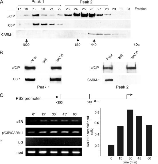
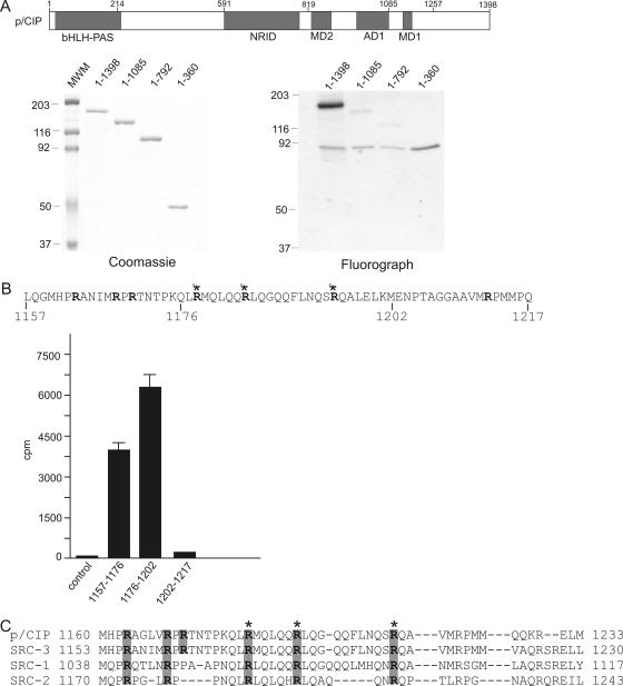
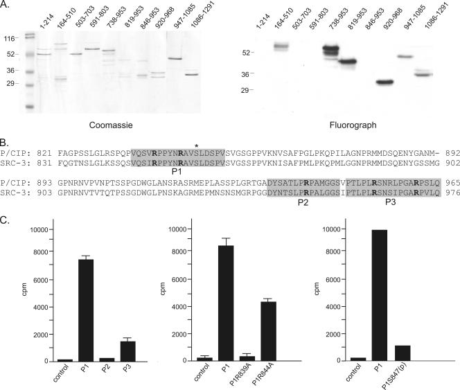
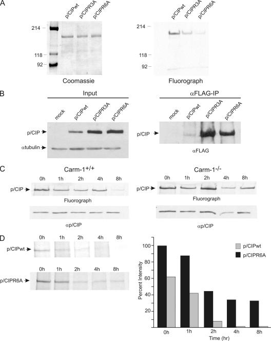
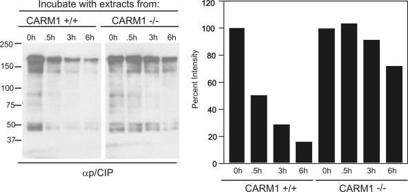
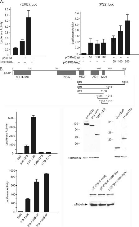
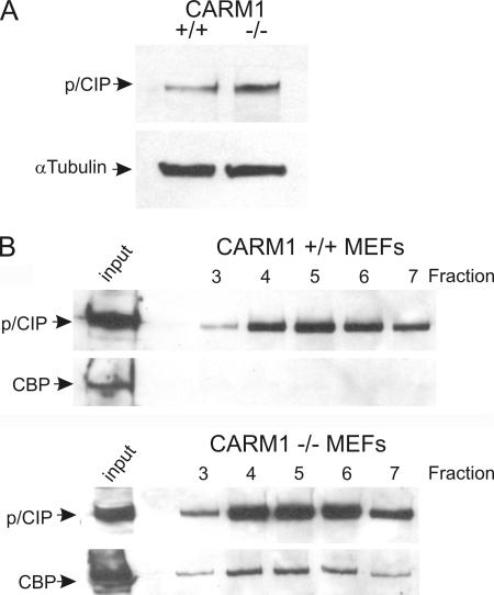
The transcriptional coactivator p/CIP(SRC-3/AIB1/ACTR/RAC3) binds liganded nuclear hormone receptors and facilitates transcription by directly recruiting accessory factors such as acetyltransferase CBP/p300 and the coactivator arginine methyltransferase CARM1. In the present study, we have established that recombinant p/CIP (p300/CBP interacting protein) is robustly methylated by CARM1 in vitro but not by other protein arginine methyltransferase family members. Metabolic labeling of MCF-7 breast cancer cells with S-adenosyl-L-[methyl-(3)H]methionine and immunoblotting using dimethyl arginine-specific antibodies demonstrated that p/CIP is specifically methylated in intact cells. In addition, methylation of full-length p/CIP is not supported by extracts derived from CARM1(-/-) mouse embryo fibroblasts, indicating that CARM1 is required for p/CIP methylation. Using mass spectrometry, we have identified three CARM1-dependent methylation sites located in a glutamine-rich region within the carboxy terminus of p/CIP which are conserved among all steroid receptor coactivator proteins. These results were confirmed by in vitro methylation of p/CIP using carboxy-terminal truncation mutants and synthetic peptides as substrates for CARM1. Analysis of methylation site mutants revealed that arginine methylation causes an increase in full-length p/CIP turnover as a result of enhanced degradation. Additionally, methylation negatively impacts transcription via a second mechanism by impairing the ability of p/CIP to associate with CBP. Collectively, our data highlight coactivator methylation as an important regulatory mechanism in hormonal signaling.


Figures

FIG. 1.
FIG. 1.
CARM1 associates with p/CIP on DNA. (A) HeLa nuclear extracts were separated by gel filtration chromatography, and fractions were assayed by immunoblotting using the antibodies indicated on the left. (B) Fractions corresponding to peaks I and II shown in panel A were pooled and passed through an anti-p/CIP immunoaffinity column or immunoglobulin G (IgG) column as control. After extensive washing, bound proteins were eluted using low pH buffer and assayed by immunoblotting using antibodies specific for p/CIP, CBP, or CARM1. (C) p/CIP and CARM1 bind to the endogenous PS2 promoter in vivo. ChIP analysis of the PS2 promoter in MCF-7 cells using antibodies against p/CIP and CARM1 demonstrates that both p/CIP and CARM1 are recruited to the PS2 promoter in response to estradiol. MCF-7 cells were stimulated for various time periods. Cells were fixed, and sequential ChIPs were performed using an anti-p/CIP antibody, followed by reimmunoprecipitation (ReChIP) using a CARM1-specific antibody. Samples were analyzed by PCR using specific primers flanking the PS2 promoter, and the relative abundance was quantified by densitometry.
FIG. 2.
FIG. 2.
In vitro methylation of p/CIP. (A) Epitope-tagged recombinant proteins were generated in Sf9 cells using baculovirus. Cells were then harvested, and proteins were immunopurified using anti-FLAG Sepharose. Approximately 500 ng of various purified proteins was then analyzed by SDS-PAGE and Coomassie blue staining. (B) Approximately 500 ng of purified recombinant CBP, p/CIP, or specific histones was incubated in the presence of 100 ng of either CARM1 or PRMT1 and [3H]SAM for 1 h. Proteins were then separated by SDS-PAGE and analyzed by fluorography. (C) In vitro methylation assays were performed using 500 ng of p/CIP, 100 ng of recombinant CARM1, and increasing concentrations of histones as indicated at the top of the panel. The reactions were terminated, proteins were separated by SDS-PAGE, and methylation of p/CIP was monitored by fluorography. BSA, bovine serum albumin.
FIG. 3.
FIG. 3.
p/CIP is methylated in intact cells. (A) Extracts from CARM1−/− MEFs do not support methylation of p/CIP in vitro. Approximately 500 ng of purified recombinant p/CIP protein was incubated with extracts derived from either CARM1+/+ or CARM1−/− MEFs in the presence of [3H]SAM. Methylation reactions were then analyzed by SDS-PAGE and fluorography. (B) MCF-7 cells were labeled for 1 h with l-[methyl-3H]methionine (50 μCi/ml) for 1 h, followed by cell lysis and immunoprecipitation of p/CIP (36). The complexes were then separated by SDS-PAGE and analyzed by fluorography. (C) MCF-7 cells were grown in the presence and absence of AdOx (20 μM) for 8 days, and p/CIP was immunoprecipitated. The immunoprecipitated complexes were separated by SDS-PAGE and probed with either an anti-p/CIP antibody (αp/CIP) or an antibody raised against dimethylated arginine 17 on histone H3 (αmR17).
FIG. 4.
FIG. 4.
Molecular mapping of CARM1-dependent methylation of p/CIP. p/CIP truncation mutants were generated using a baculovirus system and purified by immunoaffinity chromatography. Approximately 500 ng of the various truncation mutants was then tested as a substrate for CARM1 by in vitro methylation assay using 100 ng of purified CARM1. On the left is the Coomassie-stained gel of the purified proteins resolved on an 8% SDS-PAGE gel, and on the right is the corresponding fluorograph. (B) One microgram of biotinylated peptides corresponding to the regions of p/CIP indicated was used as a substrate, and the incorporation of [3H]SAM was measured by in vitro methylation in the presence of purified CARM1. The asterisk indicates the three dimethylated arginines identified by mass spectrometry. (C) Homology of MD1 within the SRC family of proteins. SRC3 corresponds to the human homologue of p/CIP. The shaded areas indicate conserved arginines, and asterisks indicate arginines identified by mass spectrometry in p/CIP.
FIG. 5.
FIG. 5.
Phosphorylation at S847 antagonizes methylation of p/CIP at R839. (A) GST recombinant proteins containing various regions of p/CIP spanning the full-length protein were generated in bacteria, purified, and used as substrates (500 ng) for purified CARM1 (100 ng) by in vitro methylation. At left is a Coomassie-stained gel of the GST fusion proteins used in the methylation assay resolved on a 12% SDS-PAGE gel, and at right is the corresponding fluorograph of the in vitro methylation reactions. (B) Comparison of the MD2 domains between p/CIP and SRC3. The asterisk corresponds to the phosphorylation site at aa 847. The shaded areas contain the methylation sites defined by in vitro methylation reactions using recombinant GST proteins. (C) Biotinylated peptides corresponding to the regions P1, P2, and P3 (shaded areas in panel B) were used as substrates, and the incorporation of [3H]SAM was measured by in vitro methylation in the presence of purified CARM1. Peptides corresponding to P1R839A and P1R844A contain substitutions of arginine to alanine. The peptide P1S847(p) is identical to P1 but contains a phosphorylated serine at aa 847.
FIG. 6.
FIG. 6.
The relative abundance of p/CIP is regulated by arginine methylation. Epitope-tagged recombinant wild-type p/CIP (wt), or p/CIP mutants containing arginine substitutions at CARM1 methylation sites (p/CIPR3A or p/CIPR6A) were generated in Sf9 cells using baculovirus. Cells were then harvested, and proteins were immunopurified using anti-FLAG-Sepharose. Approximately 1 μg of each purified protein was analyzed by SDS-PAGE and Coomassie blue staining (left) and used as substrates for in vitro CARM1-dependent methylation reactions (right). (B) COS-1 cells were transfected with expression vectors for wild-type p/CIP (wt), p/CIPR3A, or p/CIPR6A. After 24 h cells were lysed, and whole-cell extracts were prepared. Equal amounts of protein from each extract were then analyzed by Western blotting using an anti-FLAG antibody for p/CIP or anti-tubulin antibody (left). Transfected samples were immunopurified using anti-FLAG-Sepharose, followed by immunoblotting with anti-FLAG antibody (right). (C) CARM1−/− or CARM1+/+ MEFs were incubated with [35S]methionine for 40 min, followed by incubation with nonradioactive methionine-containing medium for the indicated time periods. Cells were then lysed, and p/CIP was immunoprecipitated and then subjected to SDS-PAGE and fluorography. (D) U2OS cells were transfected with either p/CIP wild-type or p/CIP R6A mutant. After 24 h, cells were incubated with [35S]methionine for 40 min and then incubated with cold methionine-containing medium for the indicated time periods. Cells were then lysed, and p/CIP was immunoprecipitated using anti-FLAG-Sepharose and then subjected to SDS-PAGE and fluorography. Samples were quantified by densitometry and expressed as a percentage of p/CIP remaining normalized to time zero.
FIG. 7.
FIG. 7.
Methylation of p/CIP is associated with increased degradation. HeLa cells were grown in the presence of AdOx for 8 days, followed by immunoprecipitation of p/CIP. Equal concentrations of p/CIP were then incubated with extracts from CARM1+/+ or CARM1−/− MEFs in the presence of SAM for the indicated time periods. Reactions were then terminated and analyzed by Western blotting using anti-p/CIP antibody. The graph on the right indicates the relative amounts of p/CIP remaining, based on densitometry.
FIG. 8.
FIG. 8.
p/CIP methylation is associated with decreased transcriptional activity. (A) COS-1 cells were transfected with expression plasmids containing ER (10 ng), full-length wild-type p/CIP (10 ng), or p/CIPR6A (10 ng) cDNAs, along with reporter plasmids (200 ng) containing three copies of an estrogen response element (ERE3Luc) or the PS2 proximal promoter (PS2Luc). Cells were stimulated with 10−6 M estradiol overnight and lysed on the following day, and luciferase activity was measured as described in Materials and Methods. (B) COS-1 cells were transfected with various regions of p/CIP fused to the Gal4 DBD along with a luciferase reporter plasmid containing five Gal4 binding sites. After 24 h, cells were lysed, and luciferase activity was measured. The corresponding Western blots shown on the right of each graph compare the relative levels of expression from the transfected Gal4 fusion cDNAs with the expression of antitubulin used as a loading control. NRID, nuclear receptor interaction domain; wt, wild type.
FIG. 9.
FIG. 9.
The association between CBP and p/CIP is decreased in CARM1−/− MEFs. (A) Whole-cell extracts were prepared from CARM1+/+ MEFs or CARM1−/− MEFs (at a similar passage number). Approximately 20 μg of protein from each extract was separated by SDS-PAGE, and Western blotting was performed using anti-p/CIP antibody or antitubulin as a control. (B) Whole-cell extracts were prepared from CARM1−/− MEFs or CARM1+/+ MEFs, and the amount of starting material in the input was normalized so that each had approximately the same amount of p/CIP based on Western blotting. p/CIP was then immunopurified by passing the protein extracts through an anti-p/CIP immunoaffinity column. After extensive washing, the bound proteins were eluted using 100 mM glycine (pH 3.0). Fractions were collected and assayed for p/CIP or CBP by SDS-PAGE followed by Western blotting.

Similar articles

Cited by

References

    1. Anafi, M., Y. F. Yang, N. A. Barlev, M. V. Govindan, S. L. Berger, T. R. Butt, and P. G. Walfish. 2000. GCN5 and ADA adaptor proteins regulate triiodothyronine/GRIP1 and SRC-1 coactivator-dependent gene activation by the human thyroid hormone receptor. Mol. Endocrinol. 14:718-732. - PubMed
    1. Anzick, S. L., J. Kononen, R. L. Walker, D. O. Azorsa, M. M. Tanner, X. Y. Guan, G. Sauter, O. P. Kallioniemi, J. M. Trent, and P. S. Meltzer. 1997. AIB1, a steroid receptor coactivator amplified in breast and ovarian cancer. Science 277:965-968. - PubMed
    1. Bedford, M. T., and S. Richard. 2005. Arginine methylation an emerging regulator of protein function. Mol. Cell 18:263-272. - PubMed
    1. Beischlag, T. V., S. Wang, D. W. Rose, J. Torchia, S. Reisz-Porszasz, K. Muhammad, W. E. Nelson, M. R. Probst, M. G. Rosenfeld, and O. Hankinson. 2002. Recruitment of the NCoA/SRC-1/p160 family of transcriptional coactivators by the aryl hydrocarbon receptor/aryl hydrocarbon receptor nuclear translocator complex. Mol. Cell. Biol. 22:4319-4333. - PMC - PubMed
    1. Brown, K., Y. Chen, T. M. Underhill, J. S. Mymyrk, and J. Torchia. 2003. The coactivator p/CIP/SRC-3 facilitates retinoic acid receptor signalling via recruitment of GCN5. J. Biol. Chem. 278:39402-39412. - PubMed

Publication types

Substances