. 2006 Oct 23;175(2):261-70.
doi: 10.1083/jcb.200605196. Epub 2006 Oct 16.

SEL1L, the homologue of yeast Hrd3p, is involved in protein dislocation from the mammalian ER

Affiliations

SEL1L, the homologue of yeast Hrd3p, is involved in protein dislocation from the mammalian ER

Britta Mueller et al. J Cell Biol. .

Abstract

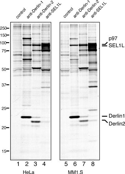
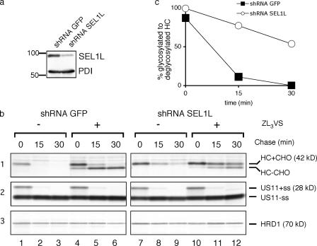
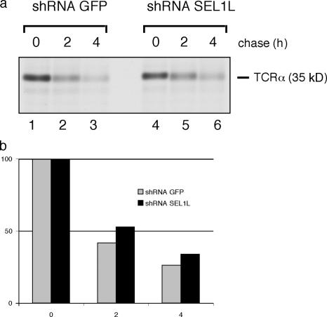
Protein quality control in the endoplasmic reticulum (ER) involves recognition of misfolded proteins and dislocation from the ER lumen into the cytosol, followed by proteasomal degradation. Viruses have co-opted this pathway to destroy proteins that are crucial for host defense. Examination of dislocation of class I major histocompatibility complex (MHC) heavy chains (HCs) catalyzed by the human cytomegalovirus (HCMV) immunoevasin US11 uncovered a conserved complex of the mammalian dislocation machinery. We analyze the contributions of a novel complex member, SEL1L, mammalian homologue of yHrd3p, to the dislocation process. Perturbation of SEL1L function discriminates between the dislocation pathways used by US11 and US2, which is a second HCMV protein that catalyzes dislocation of class I MHC HCs. Furthermore, reduction of the level of SEL1L by small hairpin RNA (shRNA) inhibits the degradation of a misfolded ribophorin fragment (RI332) independently of the presence of viral accessories. These results allow us to place SEL1L in the broader context of glycoprotein degradation, and imply the existence of multiple independent modes of extraction of misfolded substrates from the mammalian ER.


Figures

Figure 1.
Figure 1.
SEL1L is part of a conserved complex in many cell types. Immunoprecipitation from digitonin lysates of HeLa (lanes 1–4) or MM1.S (lanes 5–8) cells pulse-labeled for 16 h (steady state) were performed with control rabbit IgG, anti–Derlin-1, anti–Derlin-2, and anti-SEL1L antibodies, respectively. The positions of the relevant proteins are indicated.
Figure 2.
Figure 2.
SEL1L is an unstable ER membrane glycoprotein. Immunoprecipitation from NP-40 lysates of U373 cells pulse-labeled for 20 min and chased for the indicated time points in the absence (lanes 1–6) or presence (lanes 7–12) of ZL3VS were performed with anti-SEL1L antibodies. Half of the eluates were treated with EndoH (lanes 2, 4, 6, 8, 10, and 12).
Figure 3.
Figure 3.
shRNAs targeting SEL1L impair US11-mediated dislocation of class I MHC HC. (a) US11 cells were transduced with virus encoding shRNAs against GFP (control cells) or against SEL1L (knockdown cells) and analyzed for levels of SEL1L by immunoblotting with anti-SEL1L antibodies and anti–protein disulfide isomerase (PDI) antibodies as a loading control. (b) Control and knockdown cells were pulse-labeled for 10 min and chased for the indicated time points. Immunoprecipitation from SDS lysates for class I MHC HC (1), US11 (2), and HRD1 (3) were performed using respective antibodies. (c) Quantitation of the amount of glycosylated to deglycosylated HC from shRNA GFP (control) and shRNA SEL1L (knockdown) cells treated with ZL3VS.
Figure 4.
Figure 4.
shRNAs targeting SEL1L do not impair US2-mediated dislocation of class I MHC HC. (a) US2 cells were transduced with virus encoding shRNAs against GFP (control cells) or against SEL1L (knockdown cells) and analyzed for levels of SEL1L by immunoblotting with anti-SEL1L antibodies and anti-PDI antibodies as a loading control. (b) Control and knockdown cells were pulse-labeled for 10 min and chased for the indicated time points. Immunoprecipitation from SDS lysates for class I MHC HC (1) and US2 (2) were performed using respective antibodies. (c) Quantitation of the amount of glycosylated to deglycosylated HC from shRNA GFP (control) and shRNA SEL1L (knockdown) cells treated with ZL3VS.
Figure 5.
Figure 5.
Dislocation of truncated RI (RI332) is impaired by shRNAs targeting SEL1L. (a) HeLa cells were stably transduced with either shRNAS against GFP (control cells) or SEL1L (knockdown cells) and analyzed for levels of SEL1L by immunoblotting with anti-SEL1L antibodies and anti-PDI antibodies as a loading control. (b) Control and knockdown cells were transiently transfected with RI332. 36 h after transfection, these cells were pulse-labeled for 20 min and chased for the indicated time points. Immunoprecipitation from SDS lysates were performed using anti-RI I antibodies. (c) The bar diagram shows quantitation of the experiment. Error bars represent the SD of three independent experiments.
Figure 6.
Figure 6.
SEL1L interacts with RI332 293T cells transfected with RI332 that were pulse-labeled to steady-state for 5 h in the presence of 10 μM of the proteasome inhibitor ZL3VS. Immunoprecipitations from digitonin lysates were performed with anti-SEL1L antibodies. The anti-SEL1L immunoprecipitate was either analyzed directly (b, lane 1) or reimmunoprecipitated sequentially using anti-RI I antibodies (b, lane 2) or anti-SEL1L antibodies (b, lane 3). (a) Input levels of endogenous RI and overexpressed RI332. Immunoprecipitates were resolved on a 12% SDS polyacrylamide gel. Asterisk represents a presumably processed version of RI332 in the ER.
Figure 7.
Figure 7.
TCRα degradation is moderately slowed by shRNAs targeting SEL1L. The same control and knockdown cells used in Fig. 5 were transiently transfected with TCRα. 36 h after transfection these cells were pulse-labeled for 20 min and chased for the indicated time points. Immunoprecipitation from SDS lysates were performed using anti–TCRα-antibodies. (c) The histogram shows mean quantitation of two independent experiments.

References

    1. Ahn, K., A. Angulo, P. Ghazal, P.A. Peterson, Y. Yang, and K. Fruh. 1996. Human cytomegalovirus inhibits antigen presentation by a sequential multistep process. Proc. Natl. Acad. Sci. USA. 93:10990–10995. - PMC - PubMed
    1. Ahner, A., and J.L. Brodsky. 2004. Checkpoints in ER-associated degradation: excuse me, which way to the proteasome? Trends Cell Biol. 14:474–478. - PubMed
    1. Bays, N.W., R.G. Gardner, L.P. Seelig, C.A. Joazeiro, and R.Y. Hampton. 2001. a. Hrd1p/Der3p is a membrane-anchored ubiquitin ligase required for ER-associated degradation. Nat. Cell Biol. 3:24–29. - PubMed
    1. Bays, N.W., S.K. Wilhovsky, A. Goradia, K. Hodgkiss-Harlow, and R.Y. Hampton. 2001. b. HRD4/NPL4 is required for the proteasomal processing of ubiquitinated ER proteins. Mol. Biol. Cell. 12:4114–4128. - PMC - PubMed
    1. Bebok, Z., C. Mazzochi, S.A. King, J.S. Hong, and E.J. Sorscher. 1998. The mechanism underlying cystic fibrosis transmembrane conductance regulator transport from the endoplasmic reticulum to the proteasome includes Sec61beta and a cytosolic, deglycosylated intermediary. J. Biol. Chem. 273:29873–29878. - PubMed

Publication types

MeSH terms